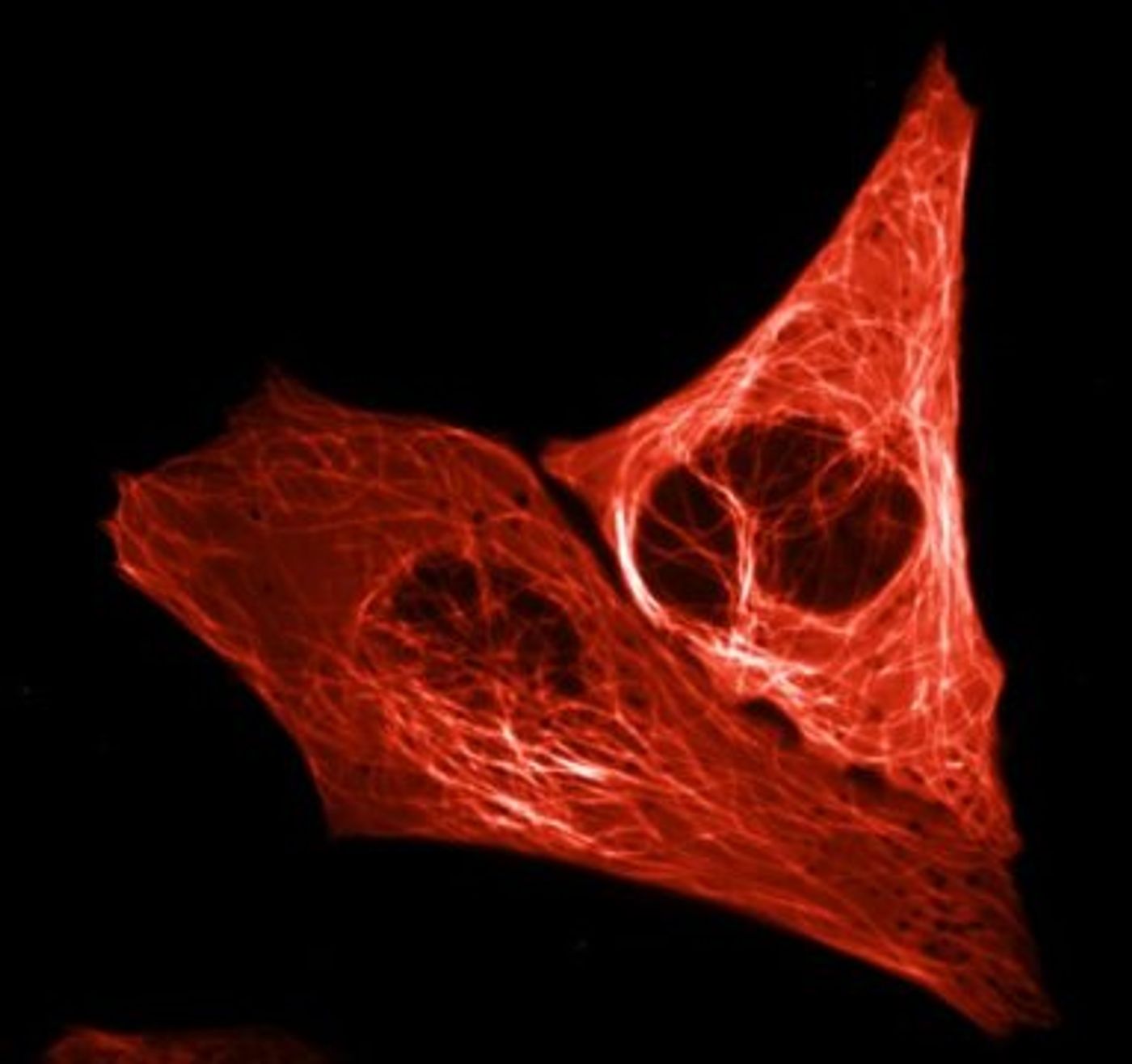
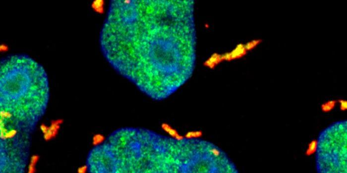
Scientists that study molecules in cells use a common tool to spotlight the proteins they are interested in - they mark stuff in the cell with tags that light up when illuminated by fluorescent lighting. It is then possible to easily and clearly see the location of proteins within a cell. One of the most common tags employed by researchers is the green fluorescent protein (GFP). It is a naturally occurring molecule that comes from fluorescent jellyfish, and has spawned many color variations over the years through manipulation of its protein code. Bright red has been an elusive color, however.
While there are some shades of red currently available, many researchers including myself will say the reds don’t match the incredible intensity of GFP.
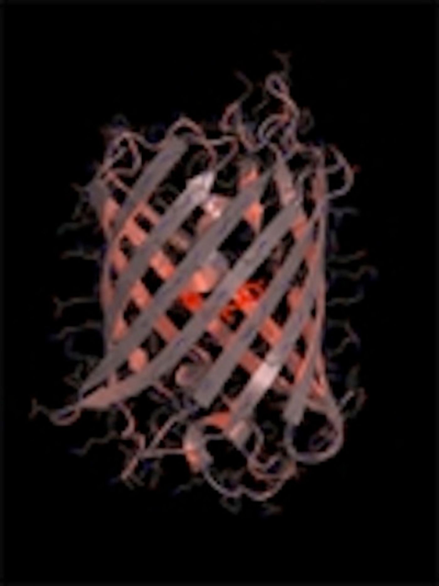
New work reported in Nature Methods by Professor of Molecular Cytology Dorus Gadella and doctoral researchers Daphne Bindels and Lindsay Haarbosch at Universiteit van Amsterdam (UVA) has produced such a red. Dubbing the new fluorescent tag mScarlet, it emits an extremely bright red shade of fluorescence. The researchers expect it to be useful for scientists working around the world in myriad fields.
The mScarlet tag was created by analyzing the varying red fluorescent proteins that have been known to exist in corals. While the corals have been seen as a chance to create a red fluorescent protein, this is the first success reported. The researchers found the genetic sequences that were common to the various coral red fluorescent proteins; deducing that sequences that were common to all of the coral would be critical parts of the protein. After putting these parts of the sequence together to make a complete strand of DNA, that whole sequence was inserted into a bacterium. The scientists then observed as it was made into a red fluorescent protein by the cellular machinery of the bacterium.
The intensity of the color was assessed, and the researchers then tinkered with the DNA code to determine how alterations in the code affected the brightness. Their experiments made for a kind of lab-based evolution in which the optimum code was determined through various methodologies.
This will hopefully be a boon to scientists studying cells under the microscope. The investigators also demonstrated that the tag does not affect proteins that it is attached to, so there seems to be no concern about unwanted side effects.
“Just as other people study the stars and prepare future trips to Mars, we are exploring the universe of the proteins that regulate the cellular processes within our bodies, “ said Gadella.
Although the research article is unfortunately behind a paywall, you can still see very short movies of the red fluorescent protein in action on the Nature Methods site - click on the videos tab under the abstract for this work
here.
Sources:
Science Daily via
UVA,
Nature Methods