Scientists at the Scripps Institution of Oceanography at the University of California, San Diego have created the Benthic Underwater Microscope, an incredible device that allows researchers to study the marine environment at a microscopic scale directly in nature. In the video below, the microscope and the work surrounding it is described and demonstrated.
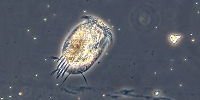
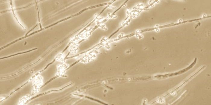
The benthic zone refers to the part of a body of water that is at the lowest level, in places where coral reefs and kelp forests grow. Studying things at a microscopic scale is, to be expected, very challenging due in part to the ever-changing nature of the ocean. Some of the images the microscope have taken are seen below.
Andrew Mullen, a Scripps Oceanography graduate student, created the microscope with colleagues and
published some of their findings in Nature Communications. They used an objective with a long working distance and electronic controls. It is capable of both single shots and time-lapse imaging and is portable enough to be carried by a diver. The scope achieves a resolution of almost a micrometer and can illuminate the working area. The instrument causes almost no disturbance to the area under study. This unique design is perfect to overcome the challenges inherent in underwater microscopy.
The researchers hope this tool can help study coral reefs, which may grow to be massive organisms, but are built by tiny little polyps, which are only about a millimeter in size. Coral reefs are currently under stress all over the world from bleaching and other problems and are considered vital parts of the marine web. As such, there is a need to know more about them.
There are many other diverse areas of research where this new scope can make an impact; studying the movements of larvae and understanding how they survive, geological particle movement on the sea floor, or how microscopic organisms compete and interact, are all processes which can now be analyzed.
Already, the Scripps research team has utilized the scope to observe battling between corals in algae in Hawaii, a fight that is ongoing. The fine scale of their documentation allowed them to learn part of the algae strategy - to start by only infringing on certain areas of the coral.
The scientists hope to usher in a wave of new research with their technology. In their paper, they even suggest a variety of upgrades that could be made to future designs. One suggestion is to add fluorescent imaging powers which would aid in the visualization of photosynthesis, or adding optical signals that indicate chemical levels. The instrument is obviously a significant improvement to the connection between the laboratory and the natural world.
Sources:
Nature Communications