
Senior Research Scientist, Molecular Devices
BIOGRAPHY


MAY 24, 2016 8:00 AM PDT
Implement high-throughput 3D image analysis for samples from subcellular structures to spheroids
Speakers
-

Steven Luke
Applications Scientist, Molecular DevicesBIOGRAPHY
Event Date & Time
DATE: May 24, 2016
TIME: 8am Pacific time, 11am Eastern time, 4pm BST, 5pm CEST
TIME: 8am Pacific time, 11am Eastern time, 4pm BST, 5pm CEST
Abstract
3D assays have been gaining popularity as researchers look for models with improved physiological relevance but scaling up these assays can be challenging in terms of creating a seamless workflow from image acquisition to analysis. In this webinar we present our latest technology advances for high-throughput imaging. Two types of assay examples will be presented:
- The development and optimization of a model system using human iPSC-derived hepatocytes to form spheroids for drug screening or assessing toxicity effects. The presentation will include experimental workflow, imaging techniques, 3D analysis and IC50 results for several compounds.
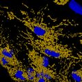
- A two-dimensional cell culture in 384-well plates treated with mitotoxic compounds. Within each cell, mitochondria can be identified and characterized to generate measurements such as average mitochondrial count, total volume, distance from the nucleus, shape factor, and intensity.
- Working with spheroids from seeding, to treating, to staining
- Imaging techniques over multiple Z planes paired with the use of our integrated, user-friendly 3D analysis toolkit
- Setting up analysis to provide data beyond intensity and number of objects, including morphology and volume of 3D structures