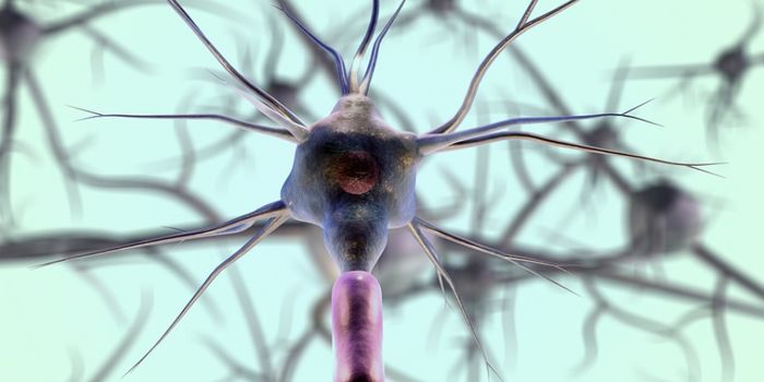
When it comes to studying the brain, imaging is everything. The technology that allows brain tissue, neurons and networks to be visualized, even at the molecular level, is constantly advancing. A new and very specific type of adaptive optics technology is being developed at Purdue University and it could be a significant improvement. The recent research by engineers at Purdue shows that high resolution images of brain cells in action can be captured in real time and might be the new way forward in understanding how the brain works. With the new process, the biology of the brain and it’s cells can be seen across a larger field of view, allowing for what the team at Purdue is calling “high throughput” imaging.

Meng Cui, an assistant professor in Purdue University’s School of Electrical and Computer Engineering and the Department of Biological Sciences explained,“We are looking at huge numbers of neurons, so the number of data points you can measure per second is 20 million, 30 million. High throughput is very important because you want to measure these numerous neurons simultaneously at very high speed and also at high spatial resolution.”
The method developed by Cui and her colleagues is called multi-pupil adaptive optics, and an important component is a deformable mirror that changes it’s configuration and shape to offset distortion that results from traditional optic imaging of tissue. Passing light through any kind of tissue will always cause a bit of distortion, but multi-pupil optics, with mirrors, creates an array of images, broken down into separate segments. Together this array of bits and pieces forms a larger field of view through the microscope.
The system is being used to look at a few specific types of brain cells. Microglia, which make up 10-15% of the cells in the brain and spinal cord are a primary focus. These are the first line of immune defense in the central nervous system. The system also looks at dendritic spines and vascular structure in the brain. Cui said in a press release, “The microglial cells are important for maintaining brain health and recovering from strokes. High-resolution in-vivo imaging of dendritic spines is also of great importance in neuroscience. And calcium imaging has been widely used in neuroscience for in-vivo large-scale recording of neuronal network activity, which demands both high speed and excellent image quality.” The lab tested the process on lab mice, detailing changes in functioning brain cells in the animals over time.
Initially the team was limited to a prism array of 3-by-3 prism array containing a total of nine segments, each around a square centimeter. That’s about 10 times better that conventional methods, but the team hopes to improve the field of view up to as many as 36 segments. The video below, from Purdue Engineering, explains more about this new method, take a look at what might be the newest way to see inside the brain.
Sources: Purdue University, Nature Methods, BioSpace