How babies develop from a fetus, to a newborn, to a toddling little human is something both scientists and parents watch with fascination. So much depends on how the brain develops, from reaching intellectual and physical milestones to interacting with the environment around them, that the infant brain has become a popular area of study. New research was recently published to give scientists a bank of information and images that perhaps someday will aid in clinical studies and even treatments for some disorders.
A large collection of MRI scans of baby brains was created as part of the Developing Human Connectome Project (dHCP). This project is a collaboration of several teams of researchers from King’s College London, Imperial College London and the University of Oxford. The project will investigate brain development in babies after they are born as well as in utero. The scans will map out brain wiring, growth and function both during pregnancy and as children grow. All the images and data collected are shared online so that researchers from around the world can access it for their own studies.
These are not just any MRI scans either. At Evelina London Children’s Hospital, the scanners are using techniques developed by the dHCP teams to address the particular difficulties of scanning babies. From keeping them still and safe in the large machines that can be loud and scary, to getting images that reveal more detail on the structure of the brain, the task is a daunting one, but one which will continue to benefit research, children and families on a global scale. The hope is that the scans can shed light on disorders like autism and how certain pregnancy complications affect fetal brain development.
Lead Principal Investigator, Professor David Edwards from King’s College London, who is also a Neonatologist Consultant at Evelina London explained, “The Developing Human Connectome Project is a major advance in understanding human brain development - it will provide the first map of how the brain’s connections develop, and how this goes wrong in disease.”
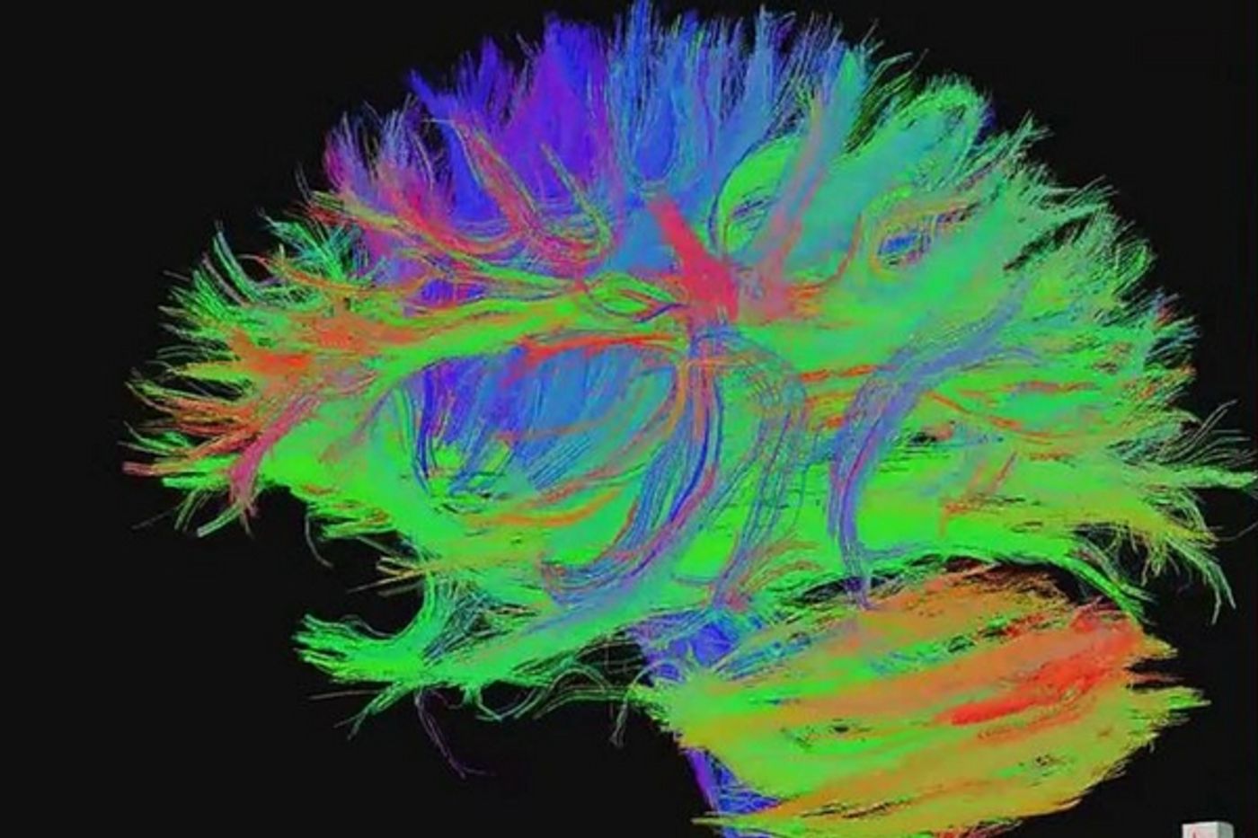
The first part of the project was to find a way to get high resolution scans, with more detail than in traditional scans. Professor Jo Hajnal at King’s College led the technology team in developing new methods that show brain structure and wiring at a level not seen before. In diffusion MRI scans, the flow of water through brain tissue was tracked and imaged to show how brain tissue grows and develops. Colleague Professor Daniel Rueckert at Imperial College London was responsible for working computer programs that can take these high-res scans and analyze what is found. Using the data to create 3D models of the brain, researchers could look at cortical surface, create sulcal depth maps, and measure mean curvature and cortical thickness. At the University of Oxford, team lead Professor Steve Smith has developed methods that map the connections in the infant brain, as these connections are a key to proper development.
The research collaboration is funded by a €15 million Synergy Grant from the European Research Council, and one of the goals of the project is to make sure that the data is shared as widely across the world as possible. The preliminary set of images can be downloaded at https://data.developingconnectome.org. The video below has more information, check it out
Sources: King’s College London, Imperial College London, Evelina London