Director, ATC Single Cell Genomics Core, Baylor College of Medicine; Professor, HGSC, Departments of Molecular and Human Genetics, Baylor College of Medicine
BIOGRAPHY

OCT 26, 2021 11:00 AM PDT
Constructing a Spatially Resolved Single-cell Atlas of the Mouse Retina with the MERSCOPE Platform
Speaker
Event Date & Time
Date: October 26, 2021
Time: 11:00am (PDT), 2:00pm (EDT)
Abstract
The retina is the light-sensing part of the visual system and is composed of six neuron types and several non-neuronal cell types, with an estimated over 100 subtypes exist based on cell morphology, function, and molecular markers. Recent single-cell transcriptomic studies show that most cell subtypes in the mouse retina can be identified based on their transcriptome. Furthermore, cell subtypes defined by their transcriptome correlated well to results obtained from morphology and function. However, a major drawback of current single-cell transcriptome technologies is that they require the isolation of single cells, resulting in the loss of spatial information. Unfortunately, spatial information is critical for studying of many biological processes, such as characterizing neuron circuit, development, and degeneration. Thus, to gain a thorough understanding of the mechanism of retinal function, construction of a spatially resolved single-cell atlas of the retina is essential. We performed single-cell spatial transcriptomic profiling on the mouse retina with MERFISH using Vizgen’s MERSCOPETM Platform. Cell organization of the retina has a distinct feature where many cell subtypes are organized in a compact area. As a result, to generate the spatial map of the retina, it is necessary to optimize the procedure. For example: 1) Due to the compactness of the retina, a new protocol needs to be developed that allows antibody staining against cell membrane along with MERFISH probes against transcripts to improve cell segmentation; 2) Cell segmentation algorithm needs to be trained and optimized for the retina; 3) As some of the cell subtypes are extremely rare (<0.01% of cell population in the retina), a novel cell annotation strategy leveraging the single-cell transcriptomic map is essential. Over 60K cells have been profiled with MERFISH. We will discuss how all cell types and most subtypes can be successfully identified and placed on the retinal spatial map.
Learning Objectives
- Identify why constructing a spatially resolved single-cell atlas of the retina is essential to gain a comprehensive understanding of retinal function
- Discover how a novel procedure was developed to perform single-cell spatial transcriptomic profiling on the mouse retina with MERFISH on Vizgen’s MERSCOPETM Platform
- Explore how MERFISH profiles and identifies cell types to enable the spatial mapping of the mouse retina
Webinars will be available for unlimited on-demand viewing after live event.
LabRoots is approved as a provider of continuing education programs in the clinical laboratory sciences by the ASCLS P.A.C.E. ® Program. By attending this webinar, you can earn 1 Continuing Education credit once you have viewed the webinar in its entirety.
You May Also Like
OCT 24, 2023 | 10:00 AM

Dynamic changes in chromatin drive gene expression programs during cellular development and contribute to pathological changes underlying disease. To date, efforts to characterize chromatin...
MAR 26, 2024 | 7:00 PM
C.E. CREDITS

The implementation of a preemptive pharmacogenomics (PGx) program in a hospital setting requires a multidisciplinary approach to ensure seamless integration of each stage of the process for...
MAR 26, 2024 | 8:00 AM
C.E. CREDITS
The implementation of a preemptive pharmacogenomics (PGx) program in a hospital setting requires a multidisciplinary approach to ensure seamless integration of each stage of the process for...
FEB 08, 2024 | 10:00 AM
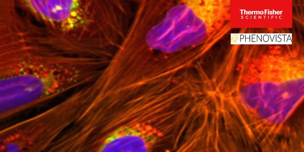
High-content screening (HCS) is an imaging-based, multi-parametric strategy used in drug development that generates rich datasets through multiplexing strategically chosen fluorescent dyes a...
MAR 26, 2024 | 11:00 AM

Ever wonder what you’re missing in your data? The sheer complexity of today’s flow and mass cytometry datasets demands automated solutions. Machine learning plugins only provide...
DEC 07, 2023 | 8:00 AM
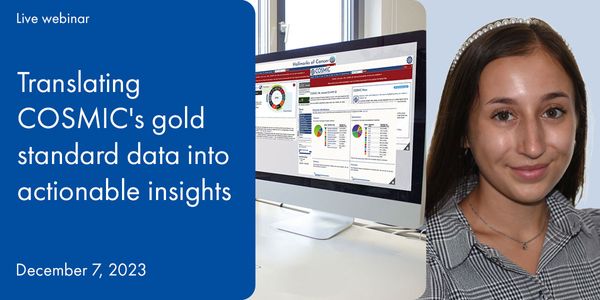
As the vast landscape of genetic oncology continues to expand, the ability to understand and utilize the full potential of this rich data becomes increasingly challenging. As a result, resea...
Loading Comments...
Finish Registering
-
 APR 30, 2024Immuno-Oncology Virtual Event Series 2024
 -
MAY 07, 20243rd International Biosecurity Virtual Symposium
-
JUN 06, 2024The Future of Scientific Conferencing
- See More


-
APR 24, 2024
- See More