Esophageal cancer remains plagued by a lack of effective techniques to detect early malignancies. Due to this major clinical challenge, esophageal cancer cases are often diagnosed at an advanced stage, with limited effective treatment options. (Our recent article covered esophageal cancer’s symptoms, treatments, and challenges in detail.)
The poor efficacy of esophageal cancer treatment methods begs the need for new techniques and approaches to screen for esophageal cancer comprehensively. A recent publication in BME Frontiers describes one such new technique that could potentially speed up our ability to detect, visualize, and diagnose esophageal cancer.
Researchers have developed a technology aptly named MAGIC (multifunctional ablative gastrointestinal imaging capsule), and their data imply a potential groundbreaking innovation to address the clinical needs surrounding esophageal cancer surveillance. MAGIC combines two necessary modalities: optical coherence tomography (OCT), a noninvasive technique for examining tissue, and tethered capsule endomicroscopy (TCE), a technology that quickly collects microscopic pictures of the esophagus in patients without sedation.
OCT uses near-infrared light to penetrate several hundred microns into a biological sample. This technology measures the amount of light which scatters off the sample and constructs a profile of the depth of the tissue at each location. Using a scanning beam of light, OCT can collect data for an entire cross-section of tissue and identify the structure. While OCT TCE technologies have shown advances in surveillance, suboptimal resolution prevents the detection of early esophageal lesions.
In developing MAGIC, researchers utilized OCT imaging at two distinct wavelengths and an ultracompact camera for TCE. The research team also added an ablation laser, which provided the capability to extract tissue for highly detailed biopsies.
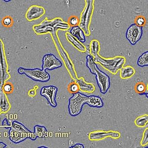
The study demonstrated the ability of MAGIC to image the esophagus in pig models. When operating at a wavelength of 800 nanometers, the researchers obtained high resolution and contrast of the surface layers of the esophagus. The images allowed the detection of early changes and abnormal growths in the esophagus. Utilizing a 1300 nanometer wavelength, the researchers could penetrate the esophageal tissue further, allowing assessment of the invasion of cancer cells between layers of tissue.
The ultracompact camera built into MAGIC helped the researchers visualize the esophagus during the procedure and provided the potential to guide laser ablation when warranted. Notably, the feasibility of MAGIC without sedation suggests a possibility of developing this technology for quick and efficient treatment of the esophagus in an outpatient setting.
MAGIC has paired complementary technologies to improve endoscopic screening and diagnosis of esophageal lesions in animal models. The authors indicate ongoing studies will continue to validate this technology and develop its clinical potential.
Sources: BME Front, J Biomed Opt, Dig Endosc