Organisms are composed of cells, some of which are constantly replaced, while others stay in place for a long time. For many years, scientists studied cell biology and tissue function by analyzing the characteristics of groups or populations of cells. But in recent years, advances in technology have enabled scientists to get a much more detailed look at specific cells by assessing them individually. Data from single-cell studies has shown that cells in a population do not always function in the same way, and has provided new insights into human health and disease. However, this research can usually only capture a snapshot of data from a cell at a specific point in time.
Now, researchers are taking the technology even further, by developing a method called Zman-seq that allows them to track cells in the body over time and follow the changes that happen in those cells. In this technique, cells are tagged with various dyes that work as time stamps, then the cells are observed in live tissue, which may be healthy or diseased. The work has been reported in Cell.
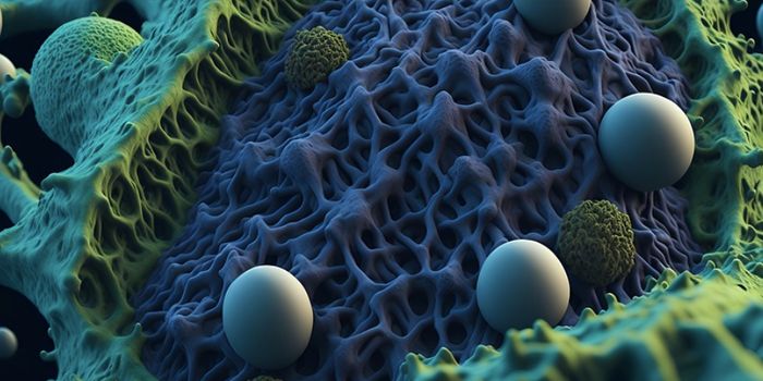
This research began with the study of aggressive brain tumors called glioblastomas. The investigators wanted to know more, not only about the rapid growth of this cancer, but also about why immune cells that get into the tumor are not able to destroy it. The scientists knew that it would be helpful to have some type of record of when an immune cell entered the tumor, and when cellular signals that would normally trigger the immune cells' activity were instead deactivated.
The investigators decided to tag specific immune cells in blood, instead of waiting until they were in the tumor tissue. They had to create different dyes that were used at different points in time, but also would not penetrate the tissue of interest, mix with one another, or hang out too long in the blood. The dyes also had to remain in place throughout the study. Once optimized, the dyes could show when a cell got into the tumor tissue, and how long it was there. The different colors could also illustrate when dysfunction arose in the immune cells. Using animal models, this approach was shown to work in the brain, lungs, and digestive systems.
The study authors were then able to use the technology to learn more about immune dysfunction in glioblastoma. They found that the mechanisms of natural killer cells, a type of immune cell that specializes in eliminating dangerous cells, are disrupted by tumors within 24 hours of entering the tumor.
"This explains why therapeutic attempts to harness the immune system for fighting glioblastoma are so ineffective," noted study co-author Dr. Daniel Kirschenbaum, a postdoctoral researcher.
Now, the scientists want to interfere with the ability of tumors to disrupt the immune system. They also want to apply Zman-seq to other types of human cells.
"For example, many cancer patients are getting therapy before surgery. We want to use the method to color immune cells in the body during that period so that after the surgery, we can better understand the dynamics of immune cells in the tumor and optimize patient treatments," added Kirschenbaum.
Experienced research scientist and technical expert with authorships on over 30 peer-reviewed publications, traveler to over 70 countries, published photographer and internationally-exhibited painter, volunteer trained in disaster-response, CPR and DV counseling.