In 2013, President Obama’s BRAIN (Brain Research Through Advancing Innovative Neurotechnologies) Initiative was launched, providing millions of dollars in funding for research into how the brain works and how new technologies can be used effectively to treat neurodegenerative diseases and traumatic brain injuries as well as mental illness and addiction. A large part of this kind of research has focused on new ways to see inside the brain, right down to the cellular level.
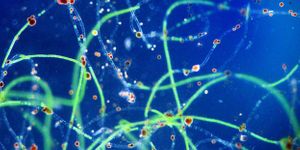
One scientist who is making progress in that area is Dr. Nancy Xu at Old Dominion University in Virginia. Her study is focused on being able to see individual neurons in the brain. The synapses between neurons, where information is exchanged are of particular interest since it’s theorized that this area is where the action that could be related to certain behaviors or illnesses begins.
Dr Xu was first awarded her grant through the EAGER (EArly-concept Grants for Exploratory Research) program of the BRAIN Initiative. Her team at Old Dominion is developing next-generation multicolored far-field photostable optical nanoscopy (PHOTON) with photostable multicolored single molecule nanoparticle optical biosensors (SMNOBS), and hopes to use this imaging platform to catch individual synapses in action and understand how individual neurons communicate with each other in real time at molecular resolution.
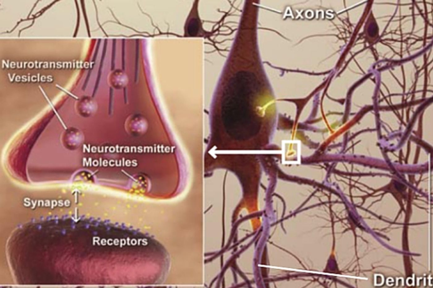
The human brain contains about 85 billion neurons and they are not all connected. Information is communicated in the brain via synapses or intersections of neurons where cell signaling is passed along pathways from neuron to neuron. Synapses are like small gaps that are connected at the cellular level when the neurons fire, sending and receiving information and passing it along through the brain. These gaps are less than 1/1000
th the width of single sheet of paper, so the interaction going on between them is incredibly difficult to visualize. The signaling of one neuron at the transmitter site and the progress of that molecular information across the synapse to the neuron receptor is an exchange that must be seen to fully understand how the brain works. If researchers could image this process, as it happens, more could be discovered about the process of disease, the causes of certain behaviors and the mechanism of addiction, to name just a few.
In a press release when she was awarded her grant, Xu stated, “Synapses enable neurons to communicate complex and specific commands with each other by passing specific electrical or chemical signals to another cell. Each synapse contains extensive arrays of molecular machinery that links the membranes of the coupled partner neurons, a wide variety of neurotransmitters and their receptors. Current tools and neurotechnologies are unable to real-time detect, image and study these wide arrays of molecular machinery and multiple types of molecules simultaneously with sufficient spatial and temporal resolutions and over an extended period of time. The technologies that we have been developing in my laboratory at ODU can potentially offer such powerful capabilities."
The video below describes the progress Dr. Xu is making on imaging at the neuronal level and where she hopes to take her project in the future. Take a look.
Sources:
Old Dominion,
National Science Foundation