A powerful new microscopy tool has been created by researchers working at the Marine Biological Laboratory (MBL) capable of taking nanoscale level measurements of the movements of individual molecules inside of living cells. This type of trafficking analysis has been extremely challenging until now. This method is even able to track the orientation of molecules.
Reporting in the Proceedings of National Academy of Sciences, the investigators describe the tool, called an "instantaneous fluorescence polarization" microscope, and how it can provide new insight into the achievement of directed functions and forces within cells.
"All functions of cells are directed. For example, cells move in a specific direction or divide at a certain site and orientation so the two daughter cells are the right size. That direction comes from the nanoscale alignment of molecules in the cells, which this microscope can detect," explained the lead author of the work, Shalin Mehta, a staff scientist at the University of Chicago's Department of Radiology and a staff researcher at MBL.
The molecules under study require visualization of really tiny particles; the action is happening on the scale of a billionth of a meter. Being able to see what is going on allows scientists to understand how complexes form from cellular components and perform necessary tasks. The technique can detect microscopic changes in conformation of proteins that are critical to functions.
"With this microscope, we can see the orientation of a single molecule, or an assembly of molecules as they form a higher-order structure," said study co-author Tomomi Tani, an MBL associate scientist.
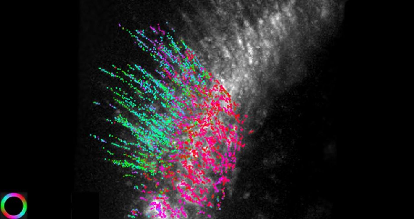
In the video above, images are shown that have been taken with the microscope. In the left panel, actin filaments of human skin have fluorescent particles move along them. On the right, the orientation of the actin filaments at the location of each particle is indicated.
Polarized light microscopes have been created and improved upon at the MBL since the 1950’s. Mehta explains that the scopes take advantage of "a property of light not visible to the human eye to measure molecular order below the resolution limit of the microscope."
This new technology has a broad set of potential applications, and can be used in vitro or in vivo. The team was able to test their microscope in various research projects with collaborators including Amy Gladfelter of University of North Carolina, Chapel Hill, and Clare Waterman of the National Institutes of Health.
"That is a unique feature of being at the MBL," Mehta commented. "We were able to study three biological questions while our method was under development. Trying to solve each question led us to improve the microscope and the algorithms with every iteration."
Sources:
Phys.org via
University of Chicago,
PNAS