Tissue engineering is a quickly growing field that involves the development of artificial organs and tissues that can be utilized to test the efficacy of drugs, repaired damaged tissues, and even to replace whole organs. However, despite its growth the field still faces challenges in current fabrication methods that limit their ability to produce free-form shapes and achieve high cell viability.
Learn more about tissue engineering:
Now, researchers at the Laboratory of Applied Photonics Devices (LAPD) in EPFL's School of Engineering, working with colleagues from Utrecht University, have developed an optical technique that takes as little as a few seconds to sculpt complex tissue shapes in a biocompatible hydrogel containing stem cells. This form of high-resolution bioprinting method, known as volumetric bioprinting, can allow the tissue to become vascularized with addition of endothelial cells.
The study was published in Advanced Materials and describes how the technique will change the way cellular engineering is performed allowing a new breed of personalized, functional bioprinted organs.
"Unlike conventional bioprinting -- a slow, layer-by-layer process -- our technique is fast and offers greater design freedom without jeopardizing the cells' viability," says Damien Loterie, an LAPD researcher and one of the study's coauthors.
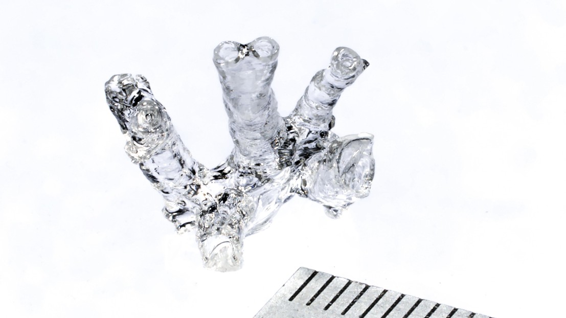
Researchers created an optical method that sculpts complex shapes in stem-cell-laden hydrogels and then vascularizes the resulting tissue to increase its viability. The groundbreaking technique will advance the field of tissue engineering. © Alain Herzog / 2019 EPFL
The first step of volumetric bioprinting involves projecting a laser down a spinning tube filled with a stem-cell-laden hydrogel. The spinning shapes the tissue by focusing the energy from the light at specific locations that then solidify. A few seconds later, a complex 3D shape appears, suspended in the gel and the stem cells present in the hydrogel are largely unaffected by this process. The endothelial cells are then added to vascularize the tissue. Researchers explain that it is possible to create a tissue construct in a clinically useful size such as a valve similar to a heart valve, a meniscus, and a part of the femur.
Bioprinting in just a few seconds:
"The characteristics of human tissue depend to a large extent on a highly sophisticated extracellular structure, and the ability to replicate this complexity could lead to a number of real clinical applications," says Paul Delrot, another coauthor.
The technique could allow mass production of artificial tissues or organs that help speed up the testing of new drugs in vitro and become ethically relevant as it obviates the need for animal testing.
"This is just the beginning. We believe that our method is inherently scalable towards mass fabrication and could be used to produce a wide range of cellular tissue models, not to mention medical devices and personalized implants," says Christophe Moser, the head of the LAPD.
Source: Science Daily