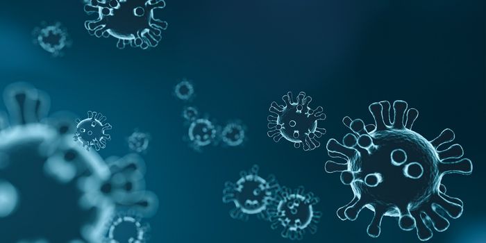
In an early release journal article from the July 2018 issue of Neoplasia, researchers sought to examine the invasive patterns of glioblastoma using invasive mouse models and clinical patients with malignant gliomas. The group found evidence that the invasive growth pattern of malignant glioma is compartmentalized; in both mice and human patients, the hippocampus was less susceptible to tumor invasion. In fact, tumor cells were visualized by MR imagery and analyzed histologically using human specific cellular markers as surrounding the hippocampal region but avoiding invasion of that boundary.
Malignant glioma infiltration patterns have been studied in the past but updates in technology and imaging have allowed these authors to design and conduct a study to further characterize those older findings. Per the authors of this study, “Early histopathological studies of brains from glioma patients showed that tumor invasion does not occur in a random manner; glioma cells follow distinct anatomical structures with a propensity to migrate along white matter tracts, in perivascular spaces and subependymal layers while avoiding certain gray matter regions.” Often, glioma subtypes are so named by their location and tendency to affect particular areas of the brain or central nervous system. It is well-known that there are multiple types of gliomas so patterns of growth and invasion as they relate to brain structures is of particular interest in this study.
Glial cells are unique and cancerous changes causing malignant growth also allow these normally large cells to change their size enough to fit in between the very small extracellular spaces within in the brain. Historical studies created malignant glioma cells through chemical manipulation or serum-cultured lines which did not necessarily act as a naturally derived invasive glioma would. This group sought to mitigate this by using patient-derived glioblastoma initiating cells (GICs) in severe combined immunocompromised mice. The literature indicates that GICs have successfully recreated the tumor environment as seen in human glioblastoma patients. Histological analysis using cellular markers demonstrated which areas of the brain were affected by cancerous cells and tumor growth. In addition to the histological analysis, the researchers confirmed malignant tumor growth through advanced MR imaging studies.
Consistent with previous studies, the invasive patterns of gliomas initially abide by anatomical borders within the brain like the pia. Even now, knowing the pia lining is a proven barrier against further spread, surgical resection of gliomas includes techniques that leave the pia intact. While some of speculated about how and why the pattern of growth and invasion is so unique, the body of knowledge surrounding this aspect of gliomas remains not well understood. The consistent finding in this study, related to invasive patterns, is the presence of non-radial migration patterns of tumor cells within the brain. Additionally, while tumor cells collected in periventricular areas, white matter tracts, and in optic and pontine white matter structures, few tumor cells invaded the hippocampal region and specific limbic structures. The authors found this finding in both mouse models and patients. It is thought that molecular signals guide glioma tumor cells to or away from certain brain compartments. Supporting this notion are the differences in migratory distances from tumor primary site or transplantation initiation site. While some primary tumor sites were located very near the hippocampus, that region was unaffected and in fact, surrounded by tumor cells that continued to invade other, more distant, tissues within the brain.
Future studies in possible immunotherapies are being explored for glioblastoma. There are encouraging results from initial phase clinical trials but not a lot of data is available as of yet. Additional research is needed to investigate glioblastoma cell marker expression, the tumor microenvironment, and how future therapies can target these very invasive cells.
Sources: Neoplasia, Johns Hopkins Health Library, National Cancer Institute, Oncoimmunology , Journal of Pathology,