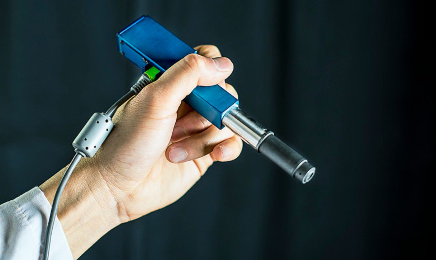
Engineers are developing a handheld microscope, roughly the size of a pen, that could help surgeons see at the cellular level where tumors stop and start.
“Surgeons don’t have a very good way of knowing when they’re done cutting out a tumor,” says Jonathan Liu, an assistant professor of mechanical engineering at the University of Washington.
“They’re using their sense of sight, their sense of touch, pre-operative images of the brain—and oftentimes it’s pretty subjective.
“Being able to zoom and see at the cellular level during the surgery would really help them to accurately differentiate between tumor and normal tissues and improve patient outcomes,” says Liu, who is senior author of a paper in
Biomedical Optics Express that describes the technology.
The microscope combines technologies in a novel way to deliver high-quality images at faster speeds than existing devices. Researchers expect to begin testing it as a cancer-screening tool in clinical settings next year.
Smaller microscopes
For instance, dentists who find a suspicious-looking lesion in a patient’s mouth often wind up cutting it out and sending it to a lab to be biopsied for oral cancer. Most come back benign.
That process subjects patients to an invasive procedure and overburdens pathology labs. Instead, physicians could use the microscope to better assess which lesions or moles are normal and which ones need to be biopsied.
“The microscope technologies that have been developed over the last couple of decades are expensive and still pretty large, about the size of a hair dryer or a small dental x-ray machine,” says study coauthor Milind Rajadhyaksha, associate faculty member in the dermatology service at the Memorial Sloan Kettering Cancer Center in New York City. “So there’s a need for creating much more miniaturized microscopes.”
Making microscopes smaller, however, usually requires sacrificing some aspect of image quality or performance such as resolution, field of view, depth, imaging contrast, or processing speed.
“We feel like this device does one of the best jobs ever—compared to existing commercial devices and previous research devices—of balancing all those tradeoffs,” says Liu.
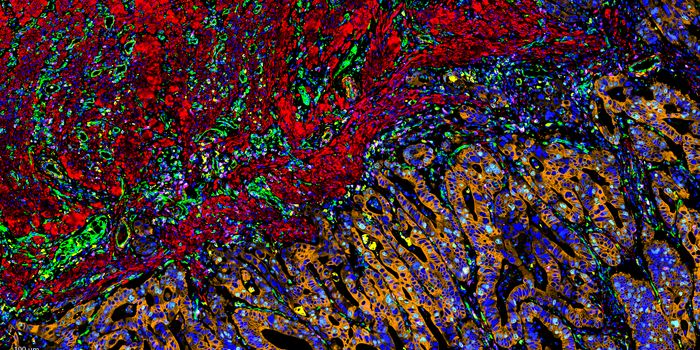
The miniature microscope uses an innovative approach called “dual-axis confocal microscopy” to illuminate and more clearly see through opaque tissue. It can capture details up to a half millimeter beneath the tissue surface, where some types of cancerous cells originate.
In the video below, for instance, researchers produced images of fluorescent blood vessels in a mouse ear at various depths ranging from 0.075 to 0.125 millimeters deep.
“Trying to see beneath the surface of tissue is like trying to drive in a thick fog with your high beams on—you really can’t see much in front of you,” Liu explains. “But there are tricks we can play to see more deeply into the fog, like a fog light that illuminates from a different angle and reduces the glare.”
The microscope also employs a technique called line scanning to speed up the image-collection process. It uses micro-electrical-mechanical—also known as MEMS—mirrors to direct an optical beam which scans the tissue, line by line, and quickly builds an image.
Imaging speed is particularly important for a handheld device, which has to contend with motion jitter from the human using it. If the imaging rate is too slow, the images will be blurry.
In the paper, the researchers demonstrate that the miniature microscope has sufficient resolution to see subcellular details. Images taken of mouse tissues compare well with those produced from a multi-day process at a clinical pathology lab—the gold standard for identifying cancerous cells in tissues.
The researchers hope that after testing the microscope’s performance as a cancer- screening tool, it can be introduced into surgeries or other clinical procedures within the next 2 to 4 years.
“For brain tumor surgery, there are often cells left behind that are invisible to the neurosurgeon. This device will really be the first to let you identify these cells during the operation and determine exactly how much further you can reduce this residual,” says project collaborator Nader Sanai, professor of neurosurgery at the Barrow Neurological Institute in Phoenix. “That’s not possible to do today.”
The National Institutes of Health is funding the project.
This article was originally published on
futurity.org.