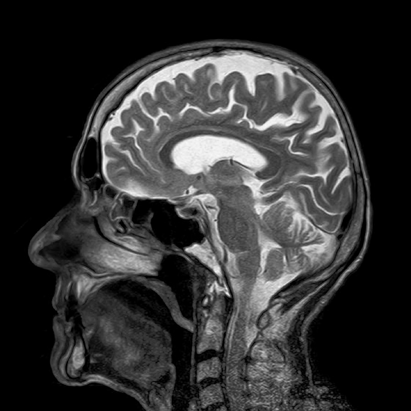
Brain imaging via MRIs may soon be enhanced with simple sugar instead of conventional metal complexes. A
pilot study found that a common form of sugar, known as dextrose, is a viable and safer alternative for tumor imaging.
Magnetic resonance imaging (MRI) is a technique that uses magnetic field and radio waves to build a picture of organs and structures inside the body. Although MRIs can be performed alone, clinicians often rely on use of agents that enhance the contrast of the image to better detect the details. An agent commonly used for this purpose is gadolinium, which is a naturally occurring metal. Gadolinium MRI contrast agents are useful, but it has known toxicities, including side effects in kidney patients. Furthermore, gadolinium can build-up in the body as deposits in the brain, especially for patients who require repeat MRIs.
To overcome this challenge, the international research team exploited tumor’s biology, mainly that tumors consume more sugar than normal tissues. Thus, they used dextrose, also known as D-glucose, as an MRI contrast agent. In a pilot study involving four normal volunteers and three patients with brain tumors, the team found comparable results between traditional gadolinium-enhanced MRIs and glucose-enhanced MRIs.
"This is the first non-metallic, biodegradable, natural MRI contrast agent tested in humans," sad Guoying Liu, Director of the National Institute of Biomedical Imaging and Bioengineering Program in Magnetic Resonance Imaging and Spectroscopy. "Developing these natural contrast agents is critical for patients who require repeated imaging; additionally it will increase patient acceptance and comfort with imaging procedures."
Using glucose could have bigger safety and efficacy potential, as noted by Peter van Zijl, the study’s senior author. According to him, recent studies have found gadolinium deposits in bone and the brain, indicating more residuals that could cause harm. “These recent published findings are showing that gadolinium deposition in tissue could be more common than previously thought, so the development of a natural contrast agent alternative is certainly a high priority for the imaging community and, of course, patients,” said van Zijl.
Furthermore, glucose may penetrate the blood-brain barrier more efficiently than gadolinium due to its smaller size. This means that glucose-enhanced MRIs should detect close to 100 percent of high-grade tumors, in contrast to the 70 to 80 percent detected by gadolinium MRIs.
"It is a significant step to be able to obtain clear MRI images of the brain using a biocompatible substance that is metabolized naturally by the body relatively quickly," said Zijl. "The dose of D-glucose is similar to that used for diabetes testing and is much cheaper than the metallic agents." In the next phase, the team will test the technique in a larger patient sample, and they the forthcoming promising results will expedite FDA approval for mass use.
Additional sources:
NIBIB