Medical imaging can offer surgeons tremendous knowledge before going into the operating room. But there’s a limit to how much a surgeon can prepare with just imaging data. In the case of a patient whose tumor was located in an especially delicate region, doctors needed addition help. They turned to 3D printing to create a model of the patient’s kidneys, which doctors used to successfully guide them through the tumor surgery before and during the operation.
The patient was Linda Green, who was diagnosed with a tumor near her kidneys about 8 years ago. Though the tumor was small in size at the beginning, it more than doubled in size since March, forcing doctors to operate. Because the tumor was located near vital components of the kidney, including its artery, veins, and ureter, Green feared the tumor removal might also mean the loss of her kidney.
But Green’s doctors had a better idea. "It's important to try to save kidneys in people every chance we can," said Jay Bishoff, director of the Intermountain Urological Institute. Together, the team at the Intermountain Medical Center in Salt Lake City decided to map out Green’s tumor and kidney structure in 3 dimension.
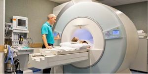
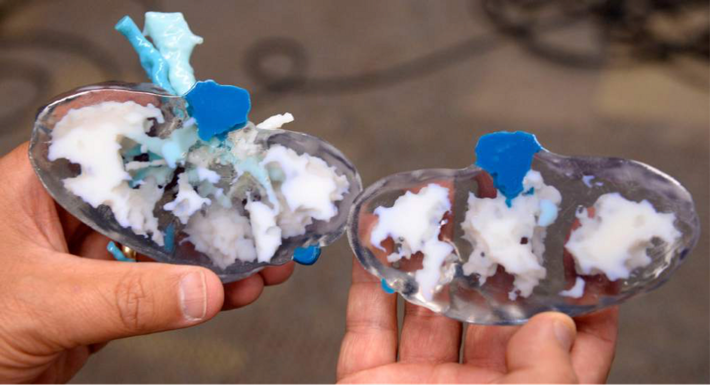
Three-dimensional printing is an innovative technique that’s rapidly gaining popularity in the biomedical arena. Using measurements gathered from in-depth computerized tomography (CT) and magnetic resonance imaging (MRI) scans, the 3D printer was able to create a true-to-form plastic model of Green’s kidney. The kidney was printed in 2 halves to show exactly how the tumor integrated into the organ. This allowed the team to plan out their exact strategies to remove the tumor without causing harm to the patient’s kidney. In studying the model, the team found a nub in the kidney that extended to where the kidney collects urine.
"Without the 3-D model, the visual images of the CT scans would not have allowed us to identify this nub prior to the surgery," said Bishoff. "The 3D printing technology allowed us to prepare a more complete plan for the patient's surgery, show the patient the complexities of the procedure and what would be done during surgery to remove the tumor and save the kidney."
The 3D printed model provided a more complete game plan than imaging scans could ever provide. In addition to studying it beforehand, the doctors also brought it into the operating room during the procedure as a reference tool.
"Dr. Bishoff was fully intent on saving my kidney, which he did and I appreciate that," said Linda. "Because he was so confident, I was confident in the fact that I was going to have a kidney. I'm just so thankful for everybody at the hospital who was involved and cared," said Green.
This is one of the few first instances where 3D printing was used to support live medical procedures. And the success of this technique may encourage doctors to utilize more 3D printed models in prepping for future medical operations in the future. "We're giving doctors additional visual tools to see the anatomy in a different way," said Cory Smith, who is part of the Transformation Lab that rendered the 3D model. "In the transformation lab we talk about reimagining imaging -- it's the evolution of imaging."
"While this technology is in its infancy, it is a big step forward in using new technologies like 3D printing to improve patient care," said Bishoff.
Additional sources:
EurekAlert!,
Salt Lake Tribune