Of the 180,000 women who have surgeries to remove cancerous breast tissues, about a quarter will return to the OR for a second time because not all the tumor was removed the first time. Now a new imaging technique has the potential to let doctors know if they’ve successfully removed all the tumor, before closing up the patient.
"Right now, we don't have a good method to assess margins during breast cancer surgeries," said Rebecca Aft, a breast cancer surgeon, who co-authored the study.
The new technology is based on photoacoustic imaging, which takes advantage of light and sound to form images. "All molecules absorb light at some wavelength," said Lihong Wan, the study’s senior author. "This is what makes photoacoustic imaging so powerful. Essentially, you can see any molecule, provided you have the ability to produce light of any wavelength. None of the other imaging technologies can do that. Ultrasound will not do that. X-rays will not do that. Light is the only tool that allows us to provide biochemical information."
So, instead of relying on later pathology exams to reveal if tumor tissue is still left, surgeons can know in the operation room just how good their cuts were.
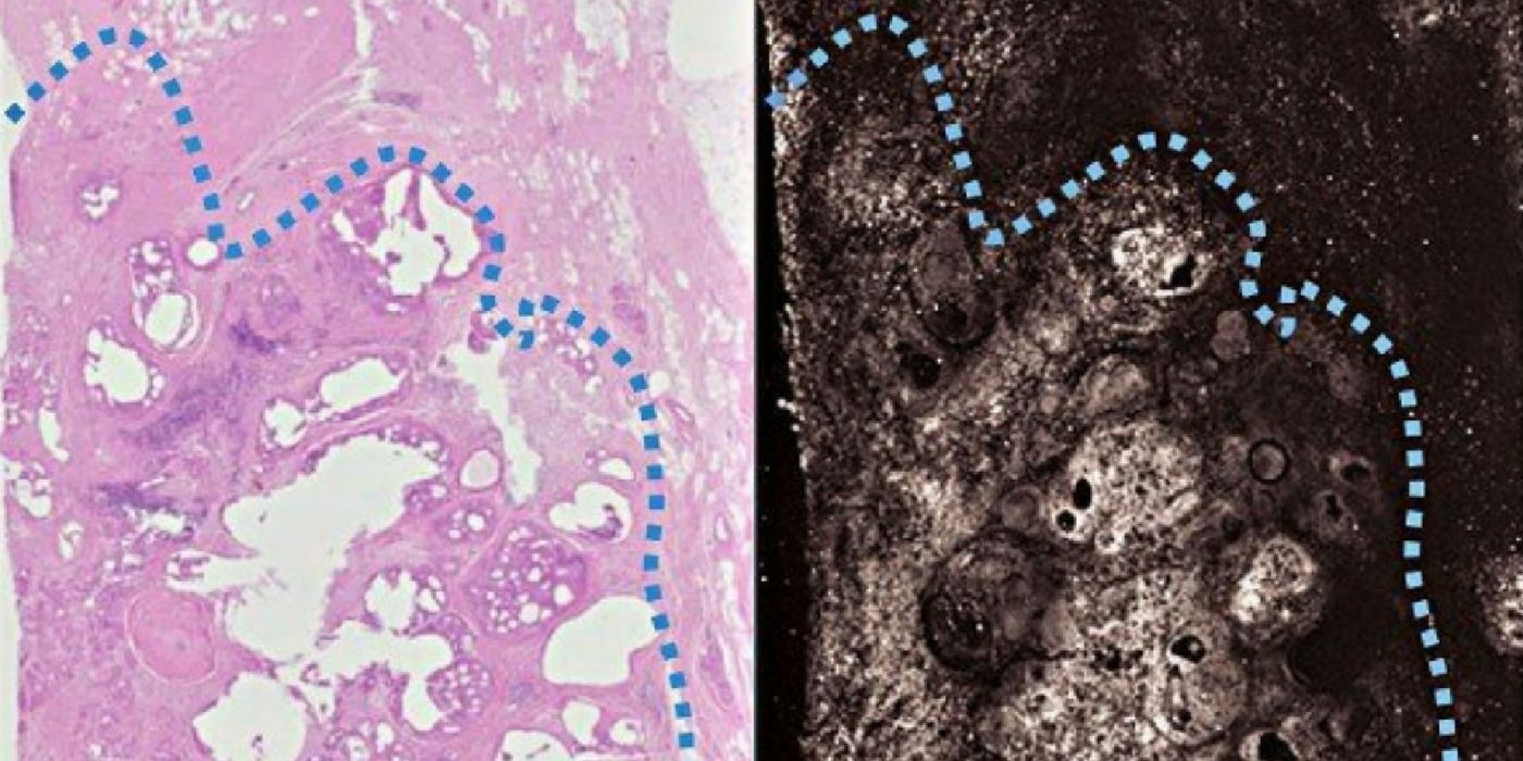
"It's the pattern of cells -- their growth pattern, their size, their relationship to one another -- that tells us if this is normal tissue or something malignant," said Deborah Novack, the study’s co-senior author. "Overall, the photoacoustic images had a lot of the same features that we see with standard staining, which means we can use the same criteria to interpret the photoacoustic imaging. We don't have to come up with new criteria."
The beauty of the photoacoustic imaging method is the speed at which information can be delivered to surgeons. With optimization, the team believes they can scan a specimen in just 10 minutes. This is vastly faster than the few days it would take a pathologist to slice, stain, and analyze the specimen under a microscope."We expect to be able to speed up the process," Wang said. "For this study, we had only a single channel for emitting light. If you have multiple channels, you can scan in parallel and that reduces the imaging time. Another way to speed it up is to fire the laser faster. Each laser pulse gives you one data point. Faster pulsing means faster data collection,” said Wang.
So instead of ending one operation only to have the patient come back for a second operation because some residual tumor was left, surgeons would be able to more precisely execute tumor removal in just one operation.
"This is a proof of concept that we can use photoacoustic imaging on breast tissue and get images that look similar to traditional staining methods without any sort of tissue processing," said Novack.
"One day we think we'll be able to take a specimen straight from the patient, plop it into the machine in the operating room and know in minutes whether we've gotten all the tumor out or not," said Aft. "That's the goal."
Additional source: Washington University in St. Louis