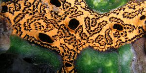
For the first time, scientists have captured on film cancer metastasis at the single-cell level in a mouse.
"One of the biggest difficulties in studying cancer," said Hiroki Ueda, the study’s co-senior author, "is that tumor metastasis is started by just a few metastasized cells. Our new method makes it possible to image the whole body down to the individual cell level, and therefore we can detect cancer at spatial resolutions beyond what is possible using other current imaging techniques."
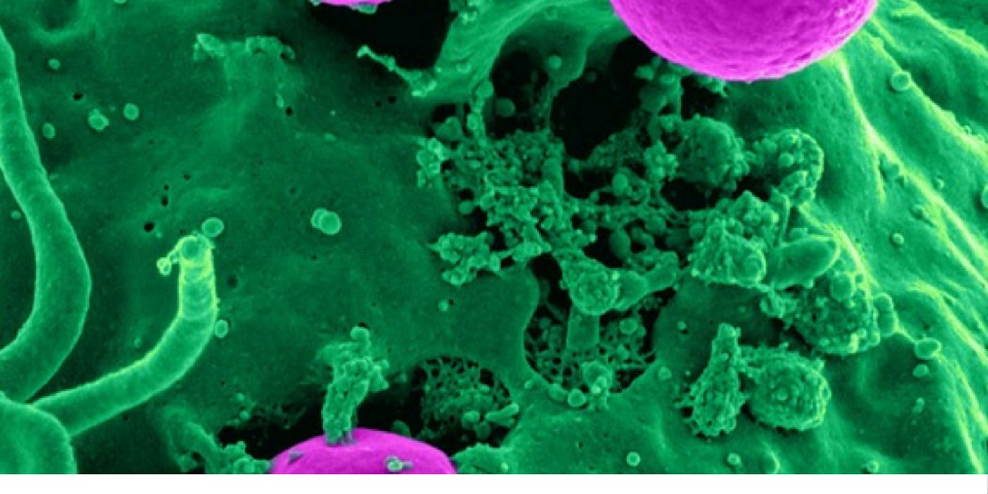
Using single-cell imaging techniques, scientists from the RIKEN Quantitative Biology Center in Japan collected data from individual cancer molecules. They combined this with a newly engineered labeling agent called CUBIC (clear unobstructed brain/body imaging cocktail).
The technique was tested on mice that were modified to have fluorescent cancer cells. Once the disease had progressed, the team applied the new imaging techniques to turn the animals’ organs transparent. This enabled the visualization of the individual cells as it multiplied and spread throughout the body.
"The images reveal cancerous colonies in enough detail to calculate their shapes, volumes, and distributions - characteristics critical to distinguishing between patterns of metastasis,” said Dr. Ueda. "We are now applying this technology to the human clinical samples. I hope this tissue-clearing and 3D imaging of human samples will make diagnosis easier, more objective and accurate in near future."
Of the current limitations and potentials of the technique, co-senior author Kohei Miyazono said, “Although we cannot apply our new CUBIC-Cancer analysis to live animals, we were able to quantify metastatic cells very early in formation. This will be a very powerful tool for evaluating the effectiveness of anti-cancer drugs.”
To demonstrate how single-cell imaging of cancer metastasis could be useful for drug screening, the team treated mice with anti-cancer drugs and surveyed the results. The technique allowed them to document differences in total volume and the number of cancer colonies in the animals’ lungs - cellular evidence of the drug’s effectiveness on cancer metastasis.
"This is very promising because these cells might be dormant or resistant to anti-cancer drugs. As just one surviving cancer cell can lead to tumor metastasis, being able to use CUBIC-Cancer analysis to evaluate drug effectiveness at this level is going to be a very useful and practical application,” said Shimpei Kubota and Kei Takahashi, the study’s co-first authors.
Additional sources: RIKEN, BBC, MNT