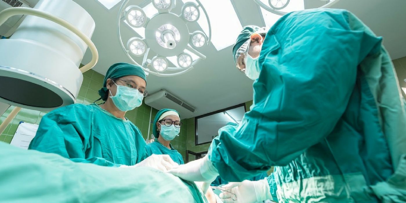
Surgical removal of the tumor remains one of the most effective lines of defense for cancer patients. However, among other drawbacks, surgery treatment is limited by how much of the tumor can be seen or felt by the surgeon. To help doctors be more thorough in the removal of ovarian tumors, scientists devised an imaging technique that penetrates deeper into the tissue, improving visualization overall.
Ovarian cancers have one of the highest mortality rates, ranking fifth in cancer deaths among women. The American Cancer Society estimated over 21,000 women in the US received a new diagnosis of ovarian cancer in 2015. Of these newly diagnosed women, about two thirds will succumb to the disease. The outcome for ovarian cancer is inherently tied to successful and complete removal of the tumor. But the removal has room for improvement, as senior study author Alexander L. Vahrmeijer explains: "Surgery is the most important treatment for ovarian cancer, and surgeons mainly have to rely on their naked eyes to identify tumor tissue, which is not optimal."
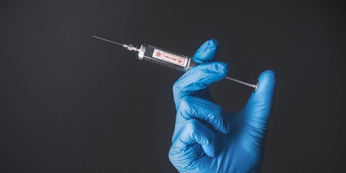
To better visualize the tumor, the research team used a new fluorescent agent called OTL38. This agent binds to a protein that’s highly expressed in ovarian cancer, marking this tissue for removal. In combination, the team devised a new imaging system known as "near infrared (NIR) fluorescence imaging.” Along with the tumor-specific marker, the NIR system allows light to penetrate deeper into tissues, in the order of centimeters.
"This allowed resection of additional tumor lesions that were not visible to the surgeons' naked eyes," said Vahrmeijer. "Although more research is needed, this is hopefully the first step toward improving the surgical outcome of cancer patients."
In clinical trials, the team tested the technique in 12 patients with ovarian cancer. They found that with the help of OTL38 and NIR, surgeons were able to remove, on average, 29 percent more malignant tissues than they could have with standard techniques.
The team plans to test this technique further in a larger study population to assess its sensitivity and specificity. "A limitation of this study is that we cannot say yet what the impact of our findings is on cure or survival of the patients. It is reasonably plausible to assume that if more cancer is removed the survival will be better. However, long-term follow-up studies need to be performed in large patient groups to prove such effects,” Vahrmeijer noted.
Additional source: American Association for Cancer Research