When graphene was first developed, it was hailed as a revolutionary material but its potential uses seemed unclear to many people. Graphene is made of a single layer of carbon atoms that form a honeycomb lattice sheet; it's extremely thin and strong. Scientists have now used a graphene camera, in which an optical image is recorded on a sheet of graphene, to record the electrical activity of a beating heart in real-time. The work has been reported in Nano Letters.
As heart muscle cells make the organ pump, their rhythmic firing generates tiny electrical fields. The voltage in neurons has traditionally been studied using stains, patch clamps, or electrodes, which usually center on a single point. But a sheet of graphene can measure the voltage in all the tissue it touches.
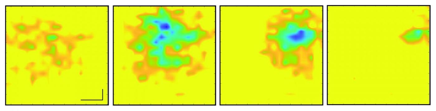
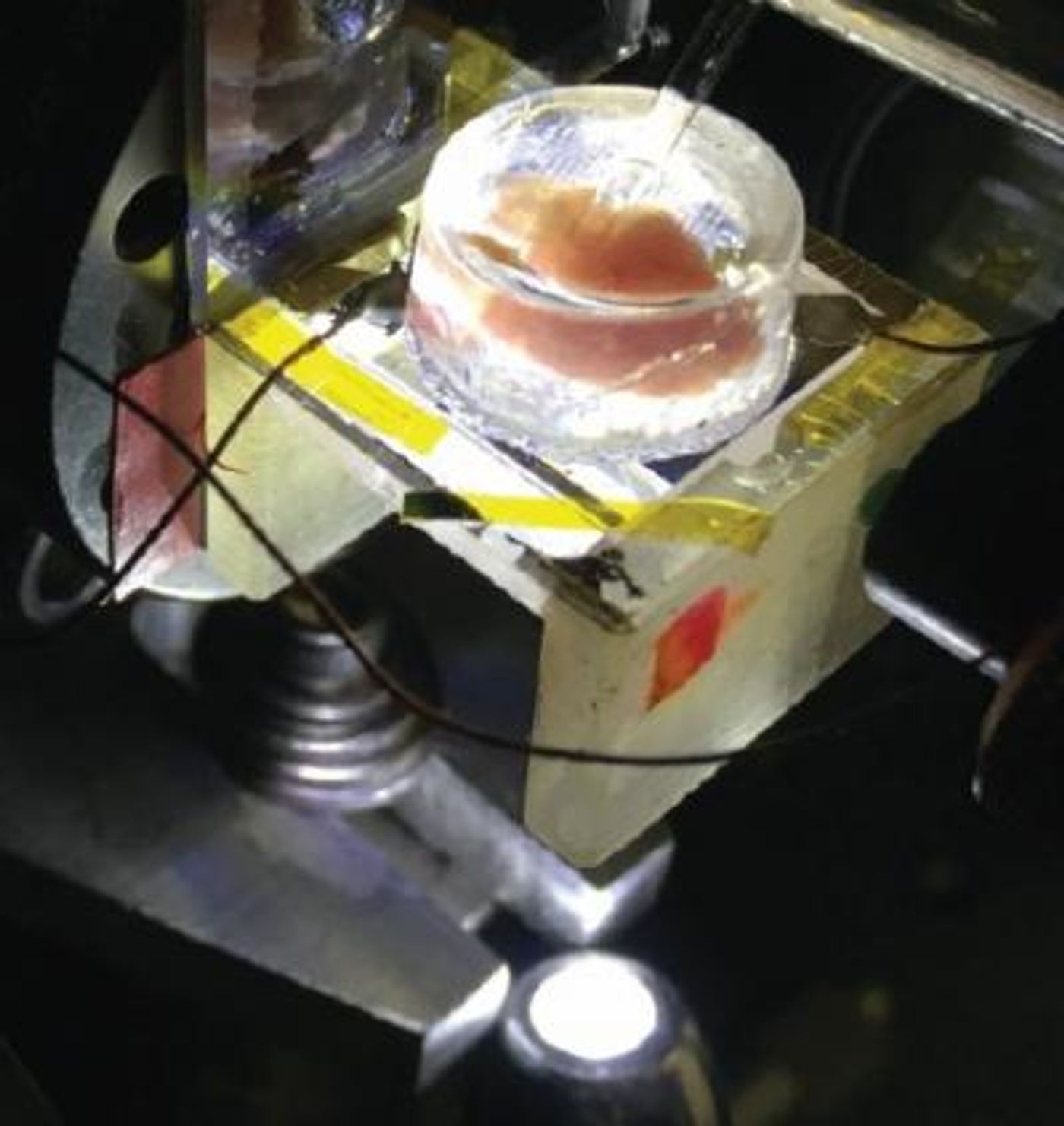
Senior study author Feng Wang, a University of California Berkeley professor of physics, discovered that electric fields can influence graphene's reflection and absorption of light. Co-first study authors Halleh Balch, Ph.D and Jason Horng, Ph.D, used the knowledge to design a graphene camera. They put a tiny sheet of graphene (about one centimeter in size) on a live chicken embryo heart and saw that the graphene sheet's reflectance changed very slightly when the electrical signals fired along the beating heart's surface.
"When cells contract, they fire action potentials that generate a small electric field outside of the cell," Balch said. "The absorption of graphene right under that cell is modified, so we will see a change in the amount of light that comes back from that position on the large area of graphene."
Since the reflectance was only changed about 2 percent by the heart's electrical field, the team amplified the signal with a waveguide under the graphene that bounced the signal internally.
"One way of thinking about it is that the more times that light bounces off of graphene as it propagates through this little cavity, the more effects that light feels from graphene's response, and that allows us to obtain very, very high sensitivity to electric fields and voltages down to microvolts," Balch explained.
Graphene sensors can sense voltage without needing tagged cells, so it can be used to record activity in nerve or muscle cells along with fluorescent stains that label other tissues.
"The ease with which you can image an entire region of a sample could be especially useful in the study of neural networks that have all sorts of cell types involved," suggested co-first study author Allister McGuire, Ph.D. "If you have a fluorescently labeled cell system, you might only be targeting a certain type of neuron. Our system would allow you to capture electrical activity in all neurons and their support cells with very high integrity, which could really impact the way that people do these network level studies."
"This is maybe the first example where you can use an optical readout of 2D materials to measure biological electrical fields," said Wang. "People have used 2D materials to do some sensing with pure electrical readout before, but this is unique in that it works with microscopy so that you can do parallel detection."
The research team, which includes quantum physicists and physical chemists from University of California, Berkeley and Stanford University called their method a critically coupled waveguide-amplified graphene electric field sensor, or CAGE sensor.
"This study is just a preliminary one; we want to showcase to biologists that there is such a tool you can use, and you can do great imaging. It has fast time resolution and great electric field sensitivity," said Horng. "Right now, it is just a prototype, but in the future, I think we can improve the device."
"One of the things that is amazing to me about this project is that electric fields mediate chemical interactions, mediate biophysical interactions; they mediate all sorts of processes in the natural world, but we never measure them. We measure current, and we measure voltage," Balch noted. "The ability to actually image electric fields gives you a look at a modality that you previously had little insight into."
Experienced research scientist and technical expert with authorships on over 30 peer-reviewed publications, traveler to over 70 countries, published photographer and internationally-exhibited painter, volunteer trained in disaster-response, CPR and DV counseling.