A new, non-invasive technique for detecting inflammation provides a unique opportunity for the early identification of atherosclerotic plaques before they rupture and cause heart disease. The technology is a new type of positron emission tomography (PET) radiotracer: gallium-68 (Ga-68)-pentixafor.
PET is a technique that measures blood flow and metabolism, offering medical professionals a source for following along with diseases progression. Ga-68-pentixafor binds to a receptor called CXCR4 on the surface of inflammatory cells present in atherosclerotic plaques, resulting in an update for doctors on the status of an individual’s blood vessel health.
Atherosclerosis develops when plaques form in the inside of the arteries due to a progressive accumulation of lipids, inflammatory cells, and connective tissue. An atherosclerotic plaque can rupture, cause blood clots, and, subsequently, cause a loss of oxygenated blood to the heart that is a main cause of heart attack and stroke. To prevent adverse cardiac events, medical professionals must be able to detect atherosclerotic plaques before they can rupture and cause blood clots.
"This new radiotracer will strongly facilitate the imaging of inflammation in atherosclerotic plaques with PET and hopefully support the early detection and treatment of atherosclerosis,” explained Fabien Hyafil, MD, PhD, from Klinikum Rechts der Isar in Germany. “Thus, preventing heart attack or stroke."
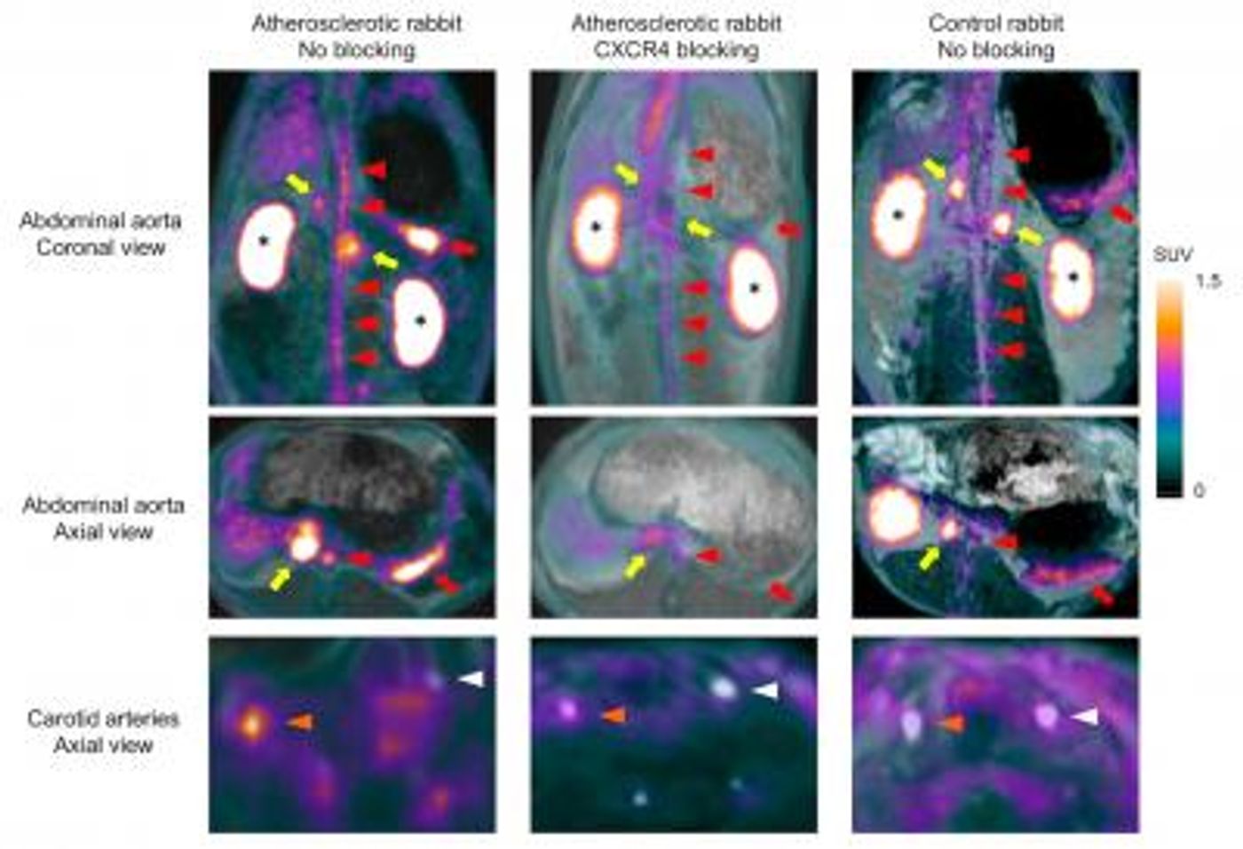
Image description: Focal activities detected with PET in atherosclerotic plaques of the abdominal aorta and the right carotid artery decreased significantly when the same rabbit was re-imaged after blocking CXCR4 receptors.
Image credit: Fabien Hyafil,MD, PhD, Department of Nuclear Medicine, Klinikum Rechts der Isar, Technische Universität München, Munich, Germany
Ga-68-pentixafor comes as a replacement for F-18-fluorodeoxyglucose (FDG)-PET, which has a few limitations. For example, patients need to fast at least six hours prior to beginning the scan process to prevent FDG from attaching to blood sugar or muscle cells that could skew the PET results. Also, FDG responds to cardiac and neural cells in addition to the desired inflammatory cells. As individuals using this technology begin to examine blood vessels closer to the heart, the scan starts to pick up signals from the cardiac cells in addition to inflammatory cells, blurring the image received depicting the presence of potential atherosclerotic plaques.
"Ga-68-pentixafor binds more specifically to inflammatory cells than FDG and does not require the patient to fast for six hours before imaging," Hyafil concluded.
Identifying potentially lethal atherosclerotic plaques before they rupture noninvasively and faster than ever before means doctors can discover and treat atherosclerosis early, preventing cardiovascular disease.
The present study was published in the Journal of Nuclear Medicine.
Sources: Society of Nuclear Medicine, National Heart, Lung, and Blood Institute, British Medical Journal