Biomedical research involves getting a close look at what’s happening inside of cells. Usually, that happens after cells are grown on a glass slide and then fixed in place. They can then be treated so that proteins or structures of interest are labeled with a color. But there are many limitations to that approach and scientists have been looking for better ways to see into cells that are still living.
A new microscope has been created that may help investigators get a better look at living cells; it uses diffraction tomography. Tomography has been utilized in many different fields, but one that’s very common is X-rays, which capture cross-sections. In light diffraction, a beam gets spread out. The technique is outlined in the video.
It's what has been employed by the HT-1, Tomocube's new microscope that produces three-dimensional images that don’t require labeled cells and can be used on living models.
These images can help researchers learn about the volume, surface area and deformability of a cell as well as giving them information about cytoplasmic density, and the shape of subcellular organelles. Not only will the information be in three dimensions it will also come from cells that have not been treated with fixatives or reagents.
“Unlike expensive atomic force, electron and laser-scanning light microscopes that require extensive sample preparation and/or labeling, the HT-1’s 3D imaging technology relies on the same fundamental property of light as traditional phase contrast microscopes,” noted Aubrey Lambert, Tomocube’s Chief Marketing Officer.
“When light passes through any object, a phase shift is observable depending on its refractive index (RI). If this emergent light is viewed together with the original light, brightness changes can be seen dependent on the degree of phase shift. In the Tomocube microscope, a 3D image, or tomogram, is created from the RI measured at each three-dimensional location during a 360-degree rotation. A similar concept using X-rays and nuclear magnetic moments is found in CT and MRI scanners,” Lambert added.
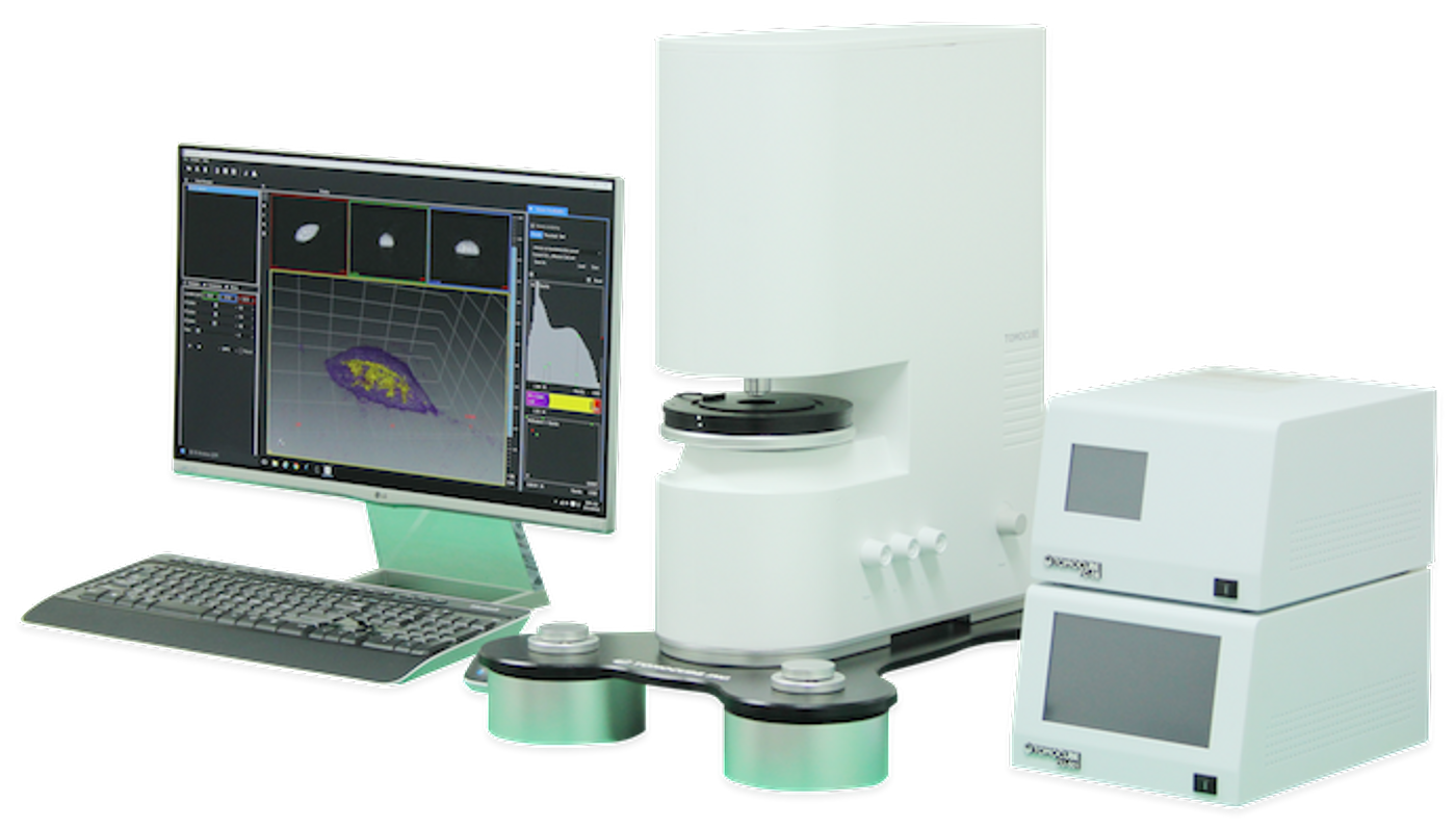
HT-1 relies on a digital micromirror device (DMD) optical light shaper, which uses hundreds of thousands of micromirrors placed on a rectangular array. Every single mirror can be tilted several degrees, and with DMD, there aren’t any moving parts interfering with the lightpath. The microscope is controlled by software created by Tomocube.
“Cellular analysis plays a crucial role in a wide variety of research and diagnostic activities in the life sciences although the information available is limited by current microscopy techniques. The Tomocube HT-1 overcomes many of these limitations and open new vistas for researchers and clinicians to understand, diagnose and treat human diseases,” said Lambert.
Source: Tomocube