For many years, clinicians have been using computed tomography (CT) scans and magnetic resonance imaging (MRI) to see inside the human body. Now scientists will be able to get a better look at cells in the brain, since a recently developed technology, three-photon microscopy, has been improved. Researchers at MIT were able to observe stimulated neurons in a live, adult mouse; in a first, they studied the layers of the visual cortex and the subplate layer below it. The findings have been reported in Nature Communications.

"By optimizing the optical design and other features for parameters for making measurements in the live brain, we were able to actually make novel discoveries that were not possible before," said co-corresponding author Mriganka Sur, Newton Professor of Neuroscience in the Picower Institute for Learning and Memory. "The concept has existed, but the question was how do you make it work.”
In this study, the researchers were able to detect patterns of activity in every layer of the visual cortex and subplate below as mice were shown images to stimulate neurons involved in their visual perception. The team showed that the technique they used did not damage or alter the cells they studied. Three-photon microscopy can deliver rapid light pulses without disturbing the cells, and the fluorescence that is emitted generates sharp images quickly.
"We were motivated to show what we could do with three-photon microscope technology for an animal in an awake condition so we could ask important questions of neuroscience," said postdoctoral fellow Murat Yildirim, a co-lead author of the study. "You could think you have the best microscope in the world, but until you ask those questions you don't know what results you are going to get."
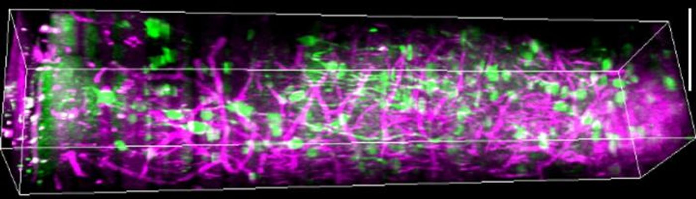
SD rendering of a sequence of 450 lateral three-photon images acquired with 2-μm increment from the visual cortex (layer 1 on the left to the subplate on the right). Green color represents GCaMP6s signal, and magenta color represents label-free THG signal generated in the blood vessels and myelin fibers in the white matter. Scale bar, 100 μm. / Credit: Murat Yildirim et. al.
The team showed mice grating patterns moving in one of two directions in a dozen different orientations. As that happened, the three-photon scope enabled them to visualize the neurons in the visual cortex - more than one millimeter deep in the brain. The neurons had been engineered to glow when they were active. The team could also see other structures like blood vessels.
The work revealed that neurons in layer five of the visual cortex could respond to a broad range of pattern orientations instead of only a few specific ones. These neurons were more spontaneously active and had more connections to deeper brain regions than cells in other layers. In layer six, the neurons were more specified in their response and only reacted to certain orientations.
Surprising the researchers, the subplate contained neurons that were tuned to visual stimuli. It had been thought that these neurons were active for the most part during development. Technical challenges, the researchers noted, have prevented the study of these cells. Now, the team will be able to use their new tool to investigate further.
Sources: AAAS/Eurekalert! via Picower Institute at MIT, Nature Communications