Studying biological processes means trying to see what is happening with molecules on the subcellular level. Visualizing these structures and proteins is a central part of biomedical research, and many scientists have devoted their efforts to improving the microscopy techniques that capture cellular functions. In recent years researchers in the lab of Ed Boyden at the Massachusetts Institute of Technology were able to create a technique that can take a tiny piece of tissue and blows it up, so tiny details are made large, and easy to see. The investigators wanted to adapt that technique to larger chunks so they could see more, like entire neuronal circuits.
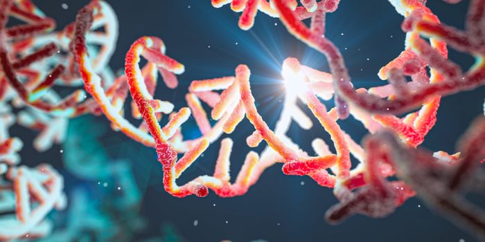
The myelin sheath acts as an insulator of nerve cells. In the mouse cortex, sheath length variation can be measured. / Credit: Gao et al./ Science 2019
Two researchers in the Boyden lab, Ruixuan Gao and Shoh Asano, turned to Howard Hughes Medical Institute investigator Eric Betzig, and his expertise in lattice light-sheet microscopy. In that technique, a very thin slice of light is shined through a tissue specimen, only illuminating the plane that is in focus, while areas that are not in the microscope’s focal plane are not subjected to intense light. That technique enables the rapid visualization of molecules inside of cells, without destroying the tissue that’s being studied. Combining expansion microscopy with the light-sheet seemed farfetched to Betzig, at first.
“I thought they were full of it,” Betzig said. “The idea does sound a bit crude,” Gao admitted. “We’re stretching tissues apart.”
Betzig still invited the researchers to take a shot. “I was going to show them,” Betzig joked. His expectations were wrong in the end. “I couldn’t believe the quality of the data I was seeing," he said. "You could have knocked me over with a feather.”
Now Boyden’s team has worked with Betzig’s group at Janelia Research Campus to capture images of all of the proteins within the whole fruit fly brain, and the mouse brain through the cortex. Generating these images with an electron microscope would take years, but it took them only 62.5 hours to image the entire fruit fly brain. The effort has been reported in Science.
“I can see us getting to the point of imaging at least ten fly brains per day,” said Betzig, who is now an HHMI investigator at the University of California, Berkeley. Working at this speed with this resolution will allow scientists to ask new questions about small details like how brain circuits in the same type of flies are different, he said.
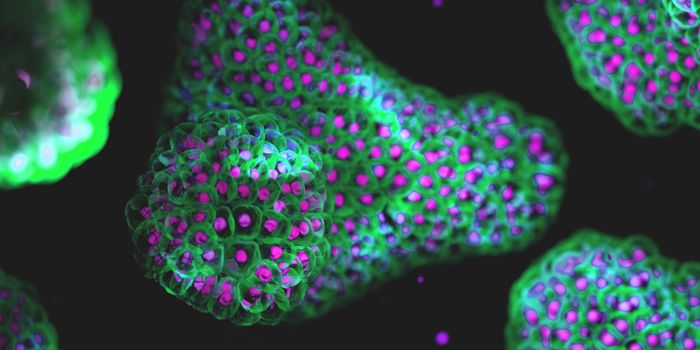
By combining expansion microscopy with lattice light-sheet microscopy, scientists can identify neuronal subsets in the fly brain and color code them by region. Credit: Gao et al./ Science 2019
Boyden’s group wants to generate maps that are detailed enough to enable computer simulations. “We’ve crossed a threshold in imaging performance,” said Boyden. “That’s why we’re so excited. We’re not just scanning incrementally more brain tissue; we’re scanning entire brains.”
There are still some limitations to this technique. Proteins must be labeled with fluorescent tags so they can be visualized by the microscope, for example. During expansion microscopy, tissues also have to be processed in many ways, which may introduce artifacts. The researchers did take the time to validate their findings in order to show that the work is reliable, however.
The team is continuing their effort to learn more about the nervous system with their light sheet technique. “Our hope is to rapidly make maps of entire nervous systems,” Boyden said.
Source: HHMI, Science