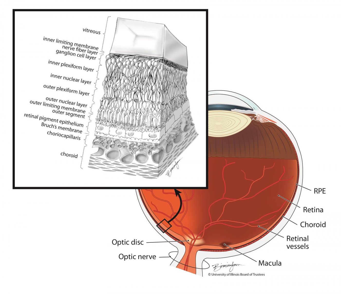
The eye contains complex layers of cells, one of which is called retinal pigment epithelium (RPE). Scientists at the National Eye Institute (NEI, a part of the National Institutes of Health) have combined a fluorescent dye with adaptive optics to observe individual cells in that critical layer. This technique can aid in the treatment of diseases that impact the RPE, like retinal degeneration. It was used in this work to compare patterns in the RPE cells of healthy individuals with patterns in retinal disease patients. The findings have been reported in JCI Insight and is summarized in the video.
"Studying cells of the retinal pigment epithelium in the clinic is like looking into a black box. RPE cells are difficult to see, and by the time signs of disease are detectable with conventional techniques, a lot of damage has often already occurred," explained the lead author of the work Johnny Tam, Ph.D. "This study is proof-of-concept that we can use a fluorescent dye to reveal this unique fingerprint of the RPE, and to monitor the tissue over time."
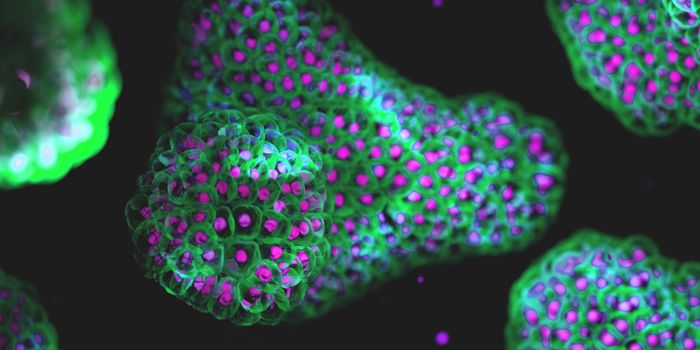
Cells in the RPE are adjacent to photoreceptors, which carry pigment and can absorb light that hits them. That means that imaging the thin RPE cell layer is challenging, even with adaptive optics. Tam decided to apply an FDA-approved dye called indocyanine green (ICG). The fluorescent dye is used to visualize blood vessels in the back of the eye and leaves those blood vessels in about thirty minutes. ICG remains in the RPE for a few hours though, and reveals a detectable pattern; some cells are dim while others are bright.
"Initially, we didn't know how the dye was going to look," said Tam. "We put the dye in, and we got this pattern that at first looked kind of random. It was a big surprise that we could come back after a year, re-inject the dye, and see the same pattern."
Tam’s team created software to assess patterns in the RPE cells and changes in those patterns, which didn’t change much in healthy people over several months.
Late-onset retinal degeneration (L-ORD) is thought to impact RPE cells in later disease stages. In an L-ORD patient in the early stages of the disease, the RPE was only a bit less stable than those in healthy people.
Bietti crystalline dystrophy (BCD) is a disease that causes the progressive loss of RPE cells. The researchers found that in a patient with BCD, RPE cells were larger and disorganized compared to healthy cells overall. There were major changes in the pattern of RPE cells impacted by BCD over time.
Tam is hopeful that the RPE patterns can be observed with conventional imaging methods eventually. That can help scientists understand dysfunction in the layer and develop ways to repair it.
"When treating patients, a lot of decisions are based on what we see. For the parts of the eye we can't see, we're often treating blindly; this technique will show us what's happening in the tissue over time, helping us develop new treatments for these conditions," concluded Tam.
Sources: AAAS/Eurekalert! Via NEI, JCI Insight