Cell division is essential to life; cells divide to build an organism and to maintain its health. The cell’s cytoskeleton, a network of filaments and microtubules inside of it, plays an important role in the process. Now researchers have revealed a novel mechanism underpinning the structure and growth of the nervous system during embryonic development, and a previously unknown role for a protein complex that plays a crucial role in cell division and has a long evolutionary history. Reporting in Developmental Cell, scientists used the common research model, Caenorhabditis elegans, to uncover new information about the coupling of microtubules and kinetochores.
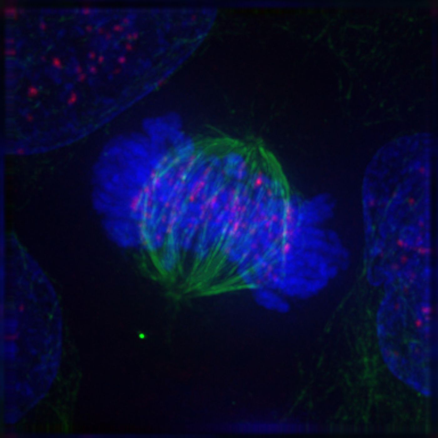
There are many stages of cell division with many details. One critical part is the duplication of the genome, which must then be distributed evenly between two new cells. Kinetochore machinery is known to play a major role in the process; it helps ensure that chromosomes are separated and end up where they're supposed to be.
In this new research, scientists have found that the machinery linking kinetochores and microtubules, the KMN network, plays an important role in the development of neurons. In the absence of the KMN network, sensory neurons don't form properly in the worm model.
"This is an entirely new discovery," said Arshad Desai, Ph.D., a professor in the Department of Cellular and Molecular Medicine at UC San Diego School of Medicine. "We've found molecular similarity between the movement of chromosomes in dividing cells and the formation of neuronal projections, both of which involve microtubule polymers that dynamically grow and shorten."
Desai is hopeful that this work will provide new insight into neurological dysfunction. Genetic mutations that impact the KMN network have been linked to a disorder in which the brain doesn’t grow properly, and the head is abnormally small, called microcephaly. "Our work suggests a potential explanation for why that happens," said Desai.
Harvard researchers have come to similar conclusions using a fruit fly model. They identified kinetochore proteins in parts of neurons of the fly, noting that a role for the kinetochore has only been found in cell division before this.
Check out an interview featuring Professor Desai in the video above.
Sources: Phys.org via UCSD, Journal of Cell Science, Developmental Cell