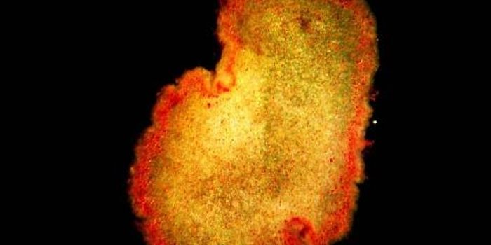
Reporting in Cancer Cell, researchers at the Walter and Eliza Hall Institute (WEHI) have created an imaging technique that enabled them to observe how cancer cells in tumors were changing. The unprecedented three-dimensional view into tumors showed how pre-cancerous cells in the mammary ducts develop into cancer over time. Breast cancer cells seem to easily morph from one type to another on the molecular level, a characteristic found in metastatic cells. Metastasis is the main cause of mortality in cancer.
Normal cells in the mammary gland can become pre-cancerous and then become early-stage cancer cells. Those cells may then change even more to become sister cells, which are individual clones descended from a tumor and are more likely to spread to other places. With their imaging tool, the researchers were able to assess how quickly pre-cancerous cells form mammary gland tumors, and observe how cells functioned there.
"Using a new imaging technique, we revealed that only a small proportion of pre-cancerous cells will develop into tumors," revealed the corresponding author of the report, Professor Jane Visvader of WEHI. "In contrast, once a tumor has been formed, we discovered it was very likely for its cells to undergo a so-called 'epithelial-to-mesenchymal transition' (EMT). This is a change in the molecular landscape - the genes that are switched on or off - within the cell, transforming it from an epithelial form to a mesenchymal form that could have a growth advantage. Our models suggest that EMT is not a rare event but is an inherent feature of mammary tumor cells."
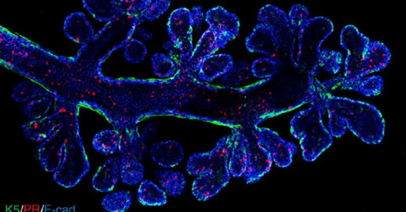
Three-dimensional view of the structure of a mammary duct. Video from Rios, Visvader et al, Cancer Cell.
These results were obtained using a research model, but the scientists believe it is close enough to human breast cancer to be applicable. They suggested that human breast cancer also carries a high level of molecular EMT.
"If EMT frequently occurs in breast cancers, it means the cells are a moving target - they can evade one set of weapons we have to fight the cancer, meaning we need to develop strategies that are more broadly targeted," said one of the researcher leaders, Professor Geoff Lindeman of WEHI.
Imaging in three dimensions was essential for this study, said Dr. Anne Rios, the first author of the report who is now at the Princess Máxima Center for Pediatric Oncology in the Netherlands.
"Until now it has been challenging to visualize the intricate structures of complex tissues such as breast tissue, or to see the true arrangement of cells within tumors," Rios added. "We developed a new, rapid way to prepare tissue samples that retains their intricate architecture but allows us to distinguish individual cells and the three-dimensional structure of the tissue.
"Our method enabled us to capture previously unseen images of breast tissue and mammary tumors - this was crucial for us to discover the frequency of EMT within the tumors," Rios said. "We expect there will be many other applications for our new imaging method, to study normal and cancerous tissue samples."
Sources: AAAS/Eurekalert! Via WEHI, Cancer Cell