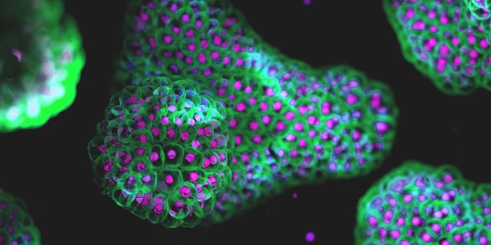
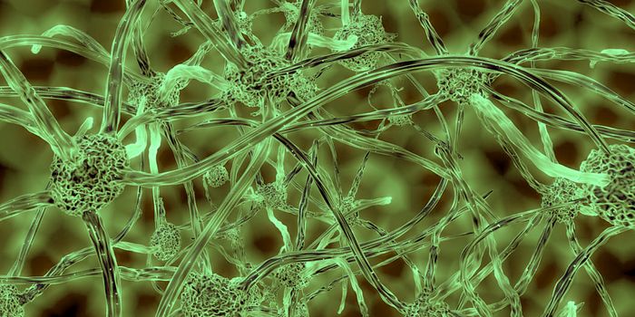
Researchers have taken powerful microscopy techniques and combined them to create a cutting edge tool for generating three-dimensional videos of individual nerve cells as they function in live fruit fly larvae that are moving at natural speeds. The team of neuroscientists and engineers have used their tool, called SCAPE, to observe proprioceptive neurons, which sense the body’s location in space, as the fly larvae are in motion. The work has been reported in Current Biology, and illustrated the function of neurons in unprecedented detail, showing how they report movement to the brain.
“We know that the brain receives sensory signals through electrical pulses passed along neurons, but we didn't understand why some kinds of neurons are located in specific positions, or how particular signaling patterns represented different movements,” explained co-senior study author and principal investigator Wesley Grueber, Ph.D. of Columbia University’s Mortimer B. Zuckerman Mind Brain Behavior Institute. "To understand this process, we needed to know what signals the neurons are sending while the larva crawled around unconstrained."
“Although we could make larvae whose neurons were labeled with fluorescence flash as they fired, we had great difficulty imaging them,” noted the first author of the report Rebecca Vaadia, a Grueber lab graduate candidate. "Even our fastest microscopes required the specimen to be constrained to move unnaturally slowly, so we could never truly capture neural activity that reflected the animal's natural, unencumbered movements until we began using SCAPE."
"We developed SCAPE to image things in 3D really, really fast," said co-senior study author and principal investigator Elizabeth Hillman, Ph.D., also of the Zuckerman Institute. "Working together, we quickly found that we could record nerve cells flashing inside fruit fly larvae as they crawled. Our high speeds allowed us to both measure the complex, 3D motions of the body and the neurons' activity during those movements. And we could do so in real time."
Because of the massive amount of data that was generated, the team wrote algorithms to track proprioceptive neurons as they activated while the body extended and compressed.
“We saw that the position of each cell made it sensitive to specific changes to the body's overall shape, and when we lined up all of the neurons' signals, we saw that they generated a detailed sequence of signals that reflected each body part's movement,” said co-first author Wenze Li, Ph.D. "It was like a beautiful machine."
“Our experiments consistently showed that each of the proprioceptor neurons reacted differently as the larvae crawled along, an observation that could not have been made if the larvae had been constrained,” said Grueber. “We saw, in real time, how some neurons fired when the animal's body stretched, while others fired when it compressed.”
The work showed that each proprioceptive neuron has its own role, sensing certain aspects of movement. This was true not only of crawling but also of more complex movement.
"We've made a lot of progress in deciphering the code of these cells," said Vaadia.
SCAPE (swept confocally aligned planar excitation), first described by Hillman’s team in 2015, scans live animals with a sheet of laser light to create 3D images. It can project and detect the sheet at the same time with one lens, generating 3D images up to 500 times quicker than other methods. It’s especially useful for small, transparent organisms, like fruit flies or zebrafish, because individual cells can be seen in a whole, live organism. Light sheet microscopy is outlined in the following video.
“Flies, worms, and fish have much simpler brains than humans, but this gives us a chance to learn how a complete nervous system actually works, cell by cell,” said Hillman. “We have good reason to believe that if we can understand these simple systems better, the lessons learned will scale up to more complex systems, including mammals.”
“We've demonstrated that SCAPE can track and map the proprioceptive neurons in a crawling fruit fly larva, and it is just the first of many studies that can now explore the fly and nervous systems of other animals,” added Li. “We can now literally label any type of cell and find out what it is doing when the animal is moving, eating or even forming a memory; the possibilities are endless.”
Sources: Phys.org via Columbia University, Current Biology