Scientists have visualized how the body uses to tests out important defensive cells and captured the process on video. The body uses immune T-cells to act as the first line of defense, monitoring the body for invading pathogens like viruses or bacteria. Before those cells are put to use, they must undergo a training process that has now been imaged and described in a report published in Nature Communications.
T-cells can also go awry, and attack the body’s own cells by mistake. That dysfunction lies at the heart of some autoimmune diseases such as type 1 diabetes, in which the insulin-producing cells of the pancreas get destroyed. This new study may offer new insights into these disorders.
"T-cells have the daunting task of recognizing and fighting off all of the diverse pathogens that we encounter throughout our lives while avoiding attacking our own healthy tissue," explained study co-author and associate professor Lauren Ehrlich. "These cells mature in the thymus, an organ just above the heart, where they 'get educated' to not attack the body."
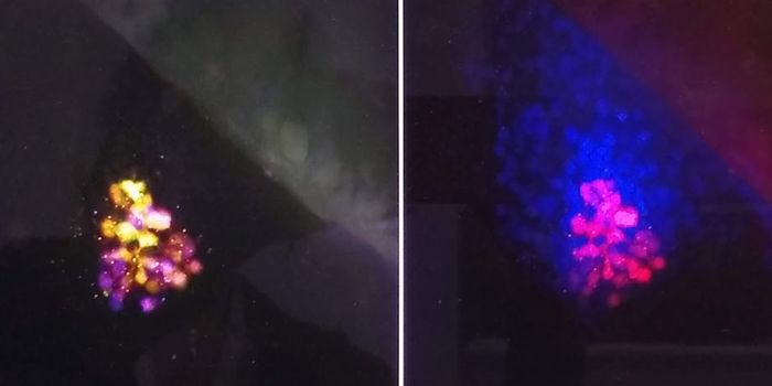
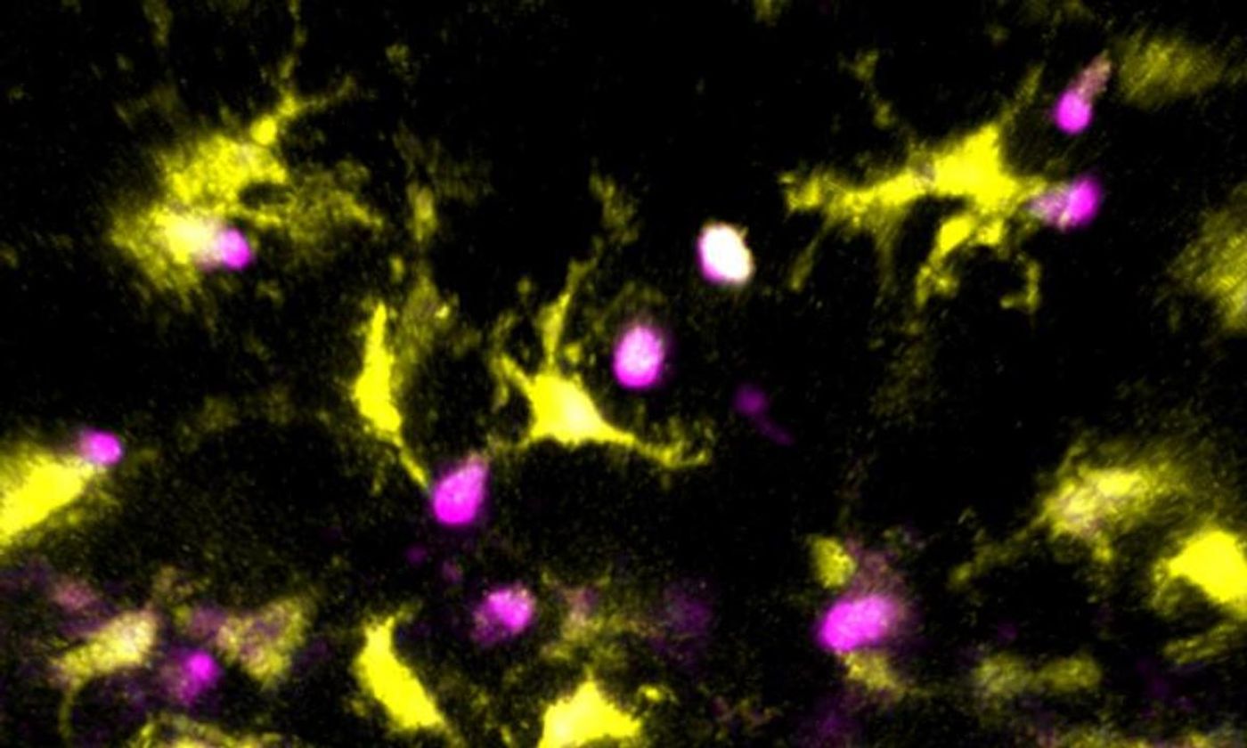
Study co-author Jessica Lancaster, a postdoctoral researcher, and Ehrlich used a mouse thymus to take video of the maturation mechanism in action. They utilized a set of powerful lasers and fired short pulses while scanning through living tissue slices. They were able to reconstruct cell signals and movements. The researchers observed other cells in the thymus helping the T-cells to mature; the T-cells were given the chance to encounter many normal human proteins that they must ignore once they are released to scan the body for invaders. Different cell types worked together to ensure that the T-cells would be safe to unleash in the body and that if they began to fail, they would self-destruct.
The researchers are hopeful that their imaging tool will help other investigators learn more about the processes occurring in the thymus.
Learn more about the maturation of T-cells in the thymus from the video.
Sources: AAAS/Eurekalert! via University of Texas at Austin, Nature Communications