The Allen Institute has developed a series of tools that can be used to explore the cell, and now they have released another. The Integrated Mitotic Stem Cell is a tool for visualizing an important type of cell division called mitosis. Cell division is critical to life; it enables organisms to grow from one cell to an entire body, and the maintenance of those bodies. This data-based work can now be used in a variety of research areas.
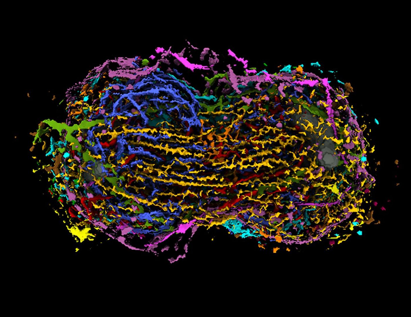
"This is the first time anyone has seen all the major parts of the cell together during mitosis," noted Rick Horwitz, Ph.D., Executive Director of the Allen Institute for Cell Science, a division of the Allen Institute. "Visualization of the data is critical. Once you see this process as a complete picture, you can start to uncover new, unexpected relationships and ask and answer completely new questions about cell division. It will also serve as a much-needed baseline of how normal human cells divide for comparison with cancer cells."
Mitosis is a carefully choreographed process, and the entire genome has to be copied and evenly divided as it occurs. Multiple proteins and organelles are involved, which work in concert as they move through quality checks and complete the production of two daughter cells. Errors can be introduced when the genome is copied, or when the genetic material is divided. Those errors can promote tumor growth.
"Mitosis is pivotal for cancer and cancer cells. To date, research on mitosis and cancer has focused mainly on chromosomes, but very few things in a cell work in isolation," said Tom Misteli, Ph.D., Director of the National Cancer Institute's Center for Cancer Research. "This new tool brings it all together, allowing researchers to connect the dots between different parts of the cell. We haven't had that until now."
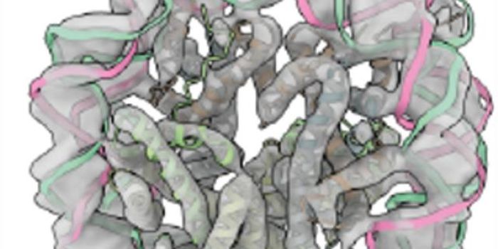
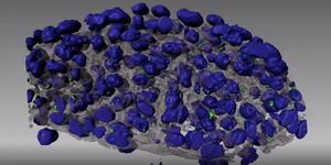
The Integrated Mitotic Stem Cell tool was created with about 400,000 images of 75 cells in the Allen Cell Collection. It provides a three-dimensional view of fifteen structures at work during cell division. It can be explored in depth in a variety of ways.
"Biologists have been looking at different images of cells and envisioning the relationships between them for decades, but to make precise comparisons, we can't just hope that everyone's mental model of a cell is the same," said Graham Johnson, Ph.D., Director of the Animated Cell team at the Allen Institute for Cell Science. "These visualizations allow us to directly look at many different structures at once by superimposing them in the same space. Now scientists can make and discuss more concrete, more accurate comparisons."
With their tool, researchers at the Allen Institute have made several observations. They found that many of the structures undergo a dramatic transformation at the same point in the early prometaphase stage of mitosis, and saw that during metaphase, the organization of the structures is at the point of lowest variance.
"These observations would not be possible without an integrated view of mitosis, but they're also just the beginning of what can be uncovered," said Susanne Rafelski, Ph.D., Director of Assay Development at the Allen Institute for Cell Science. "We hope that other researchers will find new surprises from these data that will advance our understanding of how our cells work."
Sources: AAAS/Eurekalert! via Allen Institute