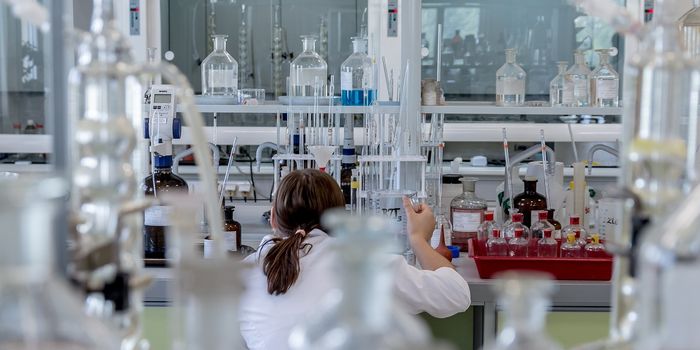
Researchers are learning more about the activity of specific nerve fibers or axons, some of which can be so long they stretch from the toes to the base of the spinal cord. Studying the role of individual axons has been challenging, but discerning their role helps us understand how neurons signal to the brain, and how the brain responds. Now a team of scientists from the Salk Institute and the University of California San Diego has developed a method for tracing the links and communications between neurons. Their work, published in Cell Reports, has revealed details of how mouse retinas receive information about light.
"This study is a breakthrough because no one could figure out how to study these connections before," noted the co-corresponding author of the study, Salk Professor Satchidananda Panda. "This new technique has enabled us to go well beyond the limitations of electron microscopy."
Several techniques were integrated for this work, which focused on neurons in the back of the eye, the retina, called intrinsically photosensitive retinal ganglion cells (ipRGCs). Those cells contain a protein called melanopsin that can sense blue light. The researchers sent a molecule called mini-singlet oxygen-generating protein (mini-SOG) into ipRGCs, enabling them to observe the action in the cells using electron microscopy. The mini-SOG was bound to the cells’ membranes, so the entire neuron, including the parts in the brain, could be visualized.
"Thanks to [the] development and application of new genetically introduced probes for correlated multiscale light and electron microscopic imaging, our Salk and UCSD-based research teams were able to follow the small processes emanating from nerve cells over centimeters, all the way from the retina to multiple places where they connect to brain regions critical to circadian rhythms, eye reflexes, and vision," explained the co-leader of the research Mark Ellisman, a distinguished professor of neurosciences at UC San Diego and adjunct professor at Salk. "We were able to obtain unprecedented three-dimensional information about the machinery required for these neuronal cells to signal the next neurons in the complex circuits."
Unlike previous work in this field that used cells grown in the laboratory, this study analyzed the cells in a mouse and revealed new details about how ipRGCs are connected to different parts of the brain. The cells were already known to link up with several areas in the brain, which play different roles. One area, for example, can control the closure of pupils in response to light, while another is involved in the regulation of the sleeping and wakefulness cycle.
"However, it takes several minutes of bright light to make us fully awake," said Panda. "How the same ipRGCs do these very different tasks with different time scales was not clear until now."
The researchers learned that information about light reaches different regions of the brain in different ways. The reaction of the pupil to light happens rapidly, they found.
"These connections were much stronger, similar to water pouring out of a garden hose," Panda said. "Whereas the connection between the ipRGCs and the master clocks were weaker, more like drip irrigation."
Since ipRGCs send signals about light to the master clock more slowly, it takes longer for information to get there and alter the clock.
"This research helps explain why, when you get up in the night to get a drink of water and turn on the light for a few seconds, you're usually able to go right back to sleep," Panda added. "But if you hear a noise outside and end up walking around your house for half an hour with the lights on, it's much harder. There will be enough light signal reaching the master clock neurons in the brain that ultimately wakes up the rest of the brain."
Their method can now be applied to the study of other neural connections, noted Panda.
"These findings and methods open new opportunities for brain researchers studying the long-distance wiring of brains in normal and in animal models of human disease," added Ellisman.
Experienced research scientist and technical expert with authorships on over 30 peer-reviewed publications, traveler to over 70 countries, published photographer and internationally-exhibited painter, volunteer trained in disaster-response, CPR and DV counseling.