Living organisms start out as one cell, and its genetic programs allow it to divide many times over, giving rise to new cells and eventually, all kinds of tissues and structures.
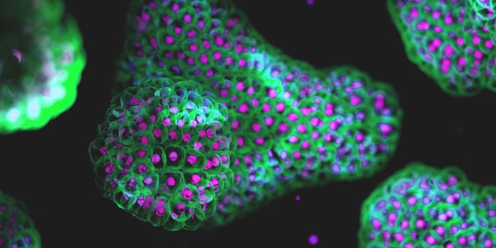
The animation above shows a 4D visualization of single-cell expression patterns. Credit: Hanna Sladitschek/EMBL
"How are the many different cell types in the body generated during embryonic development from an egg, which is only a single cell? This is one of the most fundamental questions in biology," noted Dr. Pierre Neveu, a group leader at European Molecular Biology Laboratory (EMBL) Heidelberg, as he outlined the questions his research group seeks to answer.
Assessing cellular behavior at the molecular level through all the stages of an organism's development is a challenging task, however. The activity of many genes has to be carefully orchestrated over time to ensure that development proceeds as it should, and those genes may be regulated in several different ways. Gene activity also varies depending on the cell type and developmental stage.
"So far we have lacked a comprehensive understanding of the gene expression programs. These instruct individual cells to form the different cell types necessary to build an embryo," explained the first author of the study, Dr. Hanna Sladitschek, a former EMBL Heidelberg postdoc who is now at the University of Padua School of Medicine.
In this study, the researchers constructed a virtual embryo representing a marine organism called Phallusia mammillata. This animal is a sea squirt that lives in the Atlantic Ocean and the Mediterranean Sea. Every one of these organisms has the same number of cells, so observations taken from different specimens can be combined, making an analysis like this one far easier.
The researchers used single-cell transcriptomics that shows which genes are active on the level of an individual cell, and light-sheet microscopy to image their forms as they grew from one cell to 64 during seven rounds of cell division in early development.
"Our model shows that it is possible to know the location and history of an individual cell by analyzing its gene expression," said Neveu. "In addition, we find that while the regulation of gene expression is very precise within an embryo, differences in developmental timing explain the observed variation between individual embryos."
This work illustrates that gene expression is highly coordinated during development, and is similar among many embryos, which is likely also true for other species.
"Our studies represent a leap forward in the emerging field of developmental genomics," said study co-author Dr. Lars Hufnagel. "Now that we have worked with an organism with a small number of cells, it will, of course, be very interesting to extend our work to mammals, which have many more cells!"
In the time-lapse video above, check out a zebrafish embryo developing over 24 hours from one cell to as tiny animal.
Experienced research scientist and technical expert with authorships on over 30 peer-reviewed publications, traveler to over 70 countries, published photographer and internationally-exhibited painter, volunteer trained in disaster-response, CPR and DV counseling.