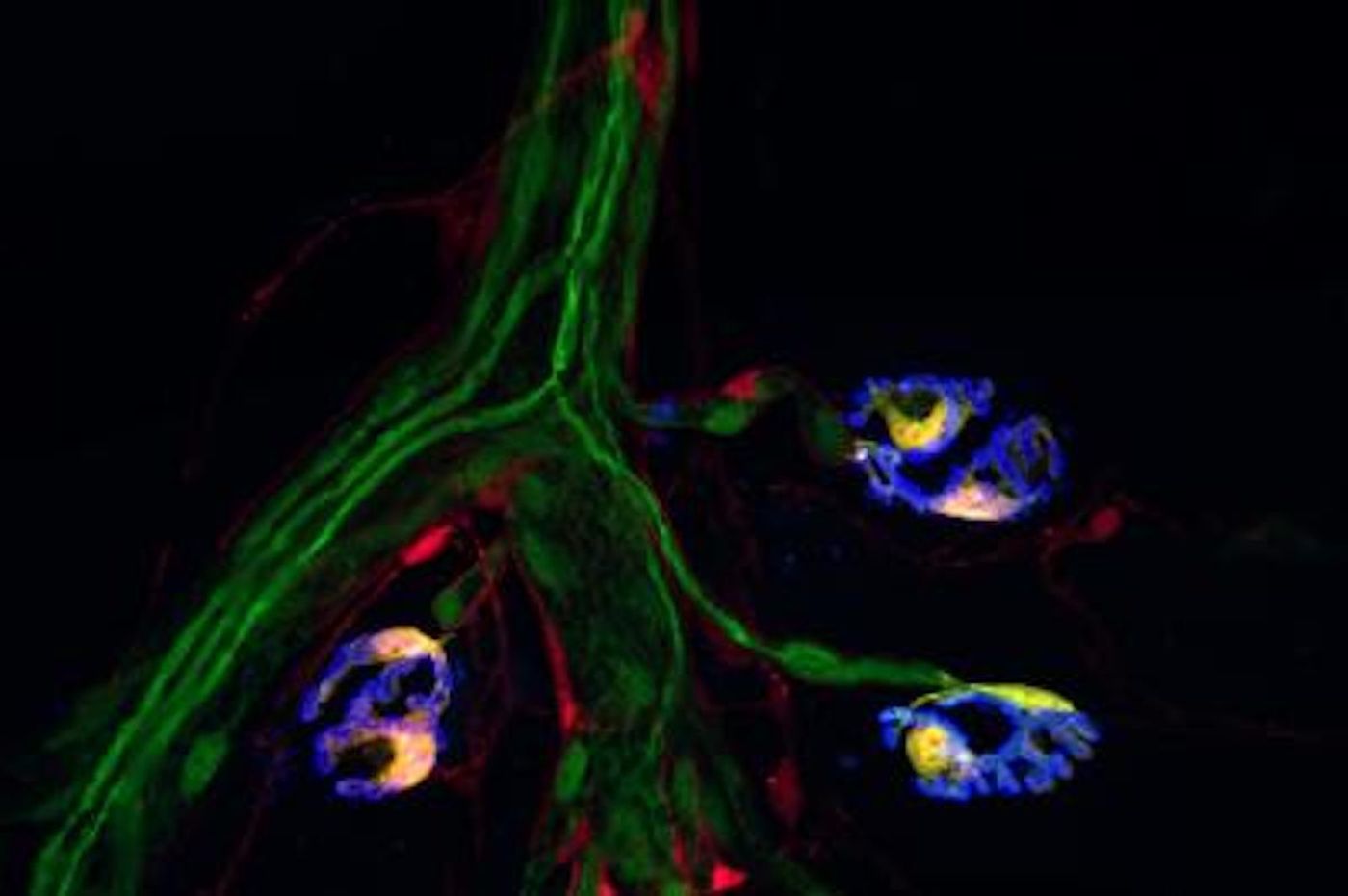
Neurons are a well-studied cell type in the brain. But another type of brain cell called glia has received less attention, even though they serve a critical function, acting as a support system for neurons. Glial cells do not send the electrical signals that neurons do, but they help ensure that they're transmitted. There are specific types of glial cells that function at the synapse - the point of contact between neurons, where biochemical signals are sent.
Scientists have now found a way to differentiate between specific kinds of glial cells, which offer new methods for studying their function. The work has been reported in eLife. It may help scientists learn more about how glial cells are involved in the restoration of nerve function after injury, aging, or disease.
"This discovery will serve as a springboard to addressing fundamental questions and developing assays to speed the discovery of therapeutics intended to preserve and restore the normal function of neuronal circuits," said the study leader Gregorio Valdez, an associate professor of molecular biology, cell biology and biochemistry at Brown University, who is affiliated with the new Center for Translational Neuroscience.
This work has shown that one type of glial cell that is found where muscle and nerve fibers meet, called Schwann cells, are the only cells in muscles that produce two specific molecules. These molecules can now act as a kind of identifier that can pinpoint this subtype of cell, said Valdez.
"What this means is that we can finally figure out how all three cellular constituents of the synapse -- neurons, muscle, and glia -- talk to each," Valdez said. "We now have a unique and important tool for identifying this critical component of the synapse. This is essential for knowing when and where to target to ensure synapses function appropriately."
This tool could prove especially useful in future work; Valdez noted that it has potential in research that investigates the pathology of amyotrophic lateral sclerosis (ALS) and spinal muscular atrophy (SMA), or how movement degenerates as we age, for example. This method may also help identify other markers for different types of glial cells.
"While our primary focus was the neuromuscular synapse, we also gathered initial evidence indicating that synaptic glia cells in the brain can be labeled and targeted using the same approach," said Valdez. "If true, this discovery could be of immense consequence for treating a myriad of brain conditions, including those involving cognitive decline due to normal aging and Alzheimer's Disease."
Experienced research scientist and technical expert with authorships on over 30 peer-reviewed publications, traveler to over 70 countries, published photographer and internationally-exhibited painter, volunteer trained in disaster-response, CPR and DV counseling.