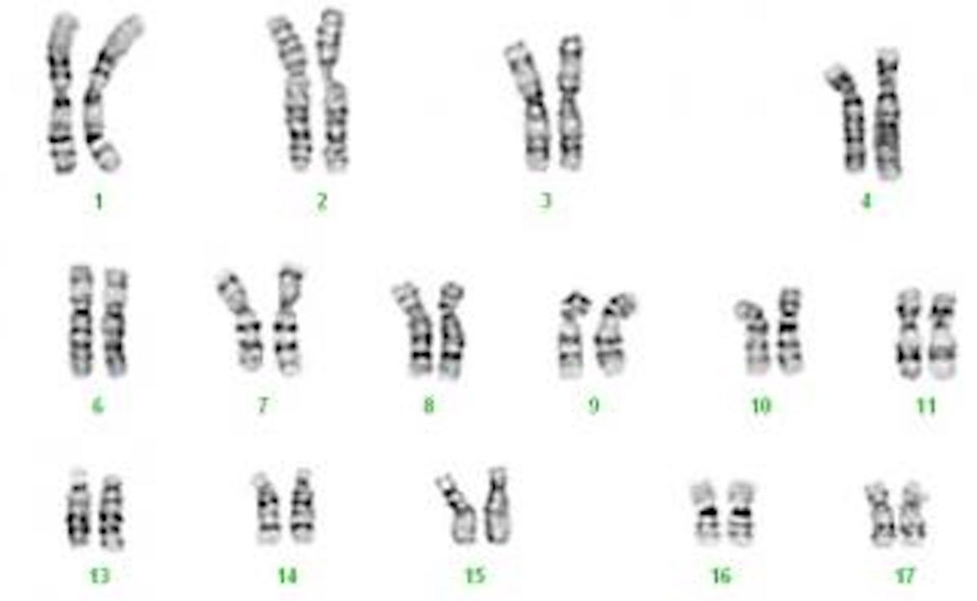
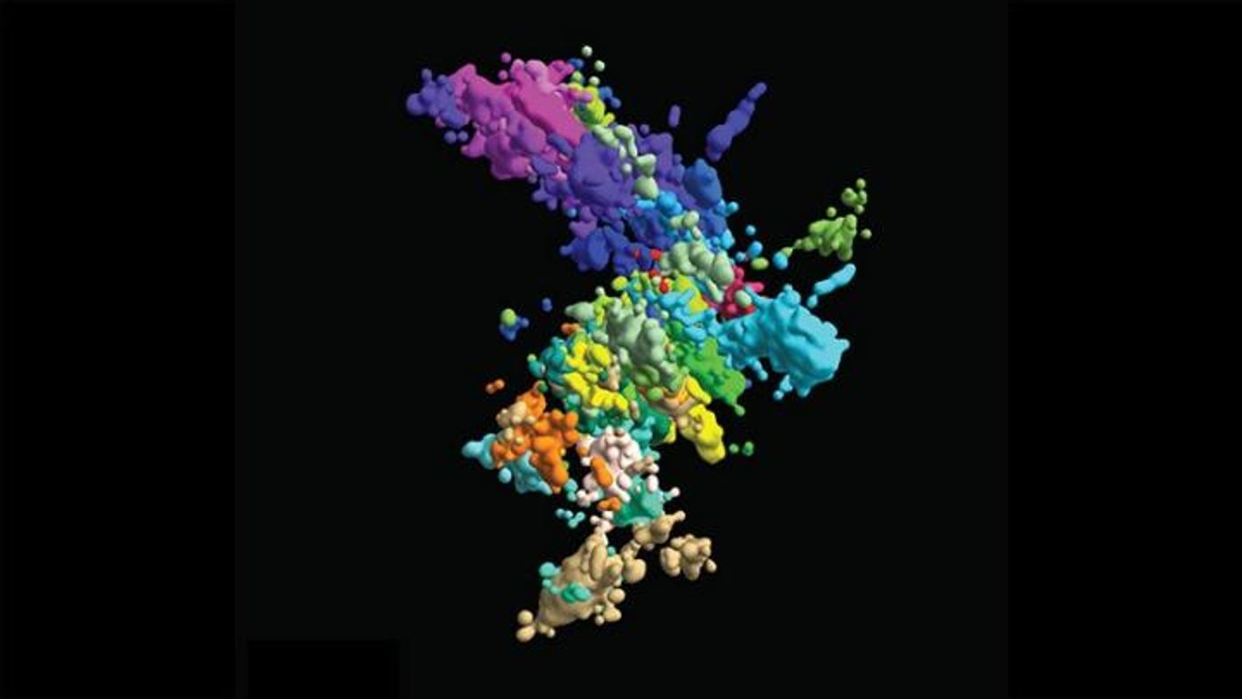
Many of us are familiar with typical diagrams of a chromosome, which is sometimes depicted like a stubby X. While that picture may resemble what is seen after some chromosome staining methods, in the cell these structures may look far different. Harvard scientists have now unveiled three-dimensional images of human chromosomes that give us a better view of reality. As it turns out, their structure is not as simple as an X-shape.
Chromosomes are DNA in its compacted form; otherwise, the genome would be far too long to fit into the nucleus of every cell. Instead, DNA has to be carefully organized and wrapped up into chromatin, which is what composes chromosomes. Chromatin is challenging to visualize, and even tougher to understand everything about how it functions.
Reporting in Cell, the team has used a novel method to capture the structure of chromatin while also imaging its behavior.
"It's quite important to determine the 3D organization to understand the molecular mechanisms underlying the organization and to also understand how this organization regulates genome function," explained Xiaowei Zhuang, the David B. Arnold, Jr. Professor of Science at Harvard University.
The technique enabled the researchers to create a chromosomal map with images of all 46 chromosomes and a close-up from one section. They did so by imaging different places on the genome, and then piecing these ‘dots’ of genomic loci together in a sequence. They had to take images of three loci at a time, cut the signal, then move on to the next three, and rapidly repeat the process.
"Now we actually have 60 loci simultaneously imaged and localized and, importantly, identified," said Zhuang.
In order to store all of this information, they used binary barcodes for different loci on the chromatin. They were able to tell the difference between 2,000 molecules in 20 imaging rounds.
"In this combinatorial way, we can increase the number of molecules that are imaged and identified much more rapidly," said Zhuang.
Since the team formed a high-resolution image of chromatin at the same time that they noted gene activity and the presence of nuclear structures like nucleoli, they could study how the structure changed over time, like during cell division or transcription.
This effort revealed that places on chromatin that contain lots of genes tend to attract other areas that are similar. But places with few genes will only connect if they are located on the same chromosome. It may be that it’s efficient for so-called gene-rich areas to be close to one another so that the cell's transcription machinery can reach it all easily.
While more work will be needed to learn why ‘like attracts like’ on chromatin, the study authors suggested that chromatin structure is affecting function.
They also learned that chromosomes look unique; it will take a very long time, and more labs, to determine what chromosomes look like in different human cells.
"It's not going to be possible to build just on our work," Zhuang said. "We need to build on many, many labs' work in order to have a comprehensive understanding."
Experienced research scientist and technical expert with authorships on over 30 peer-reviewed publications, traveler to over 70 countries, published photographer and internationally-exhibited painter, volunteer trained in disaster-response, CPR and DV counseling.