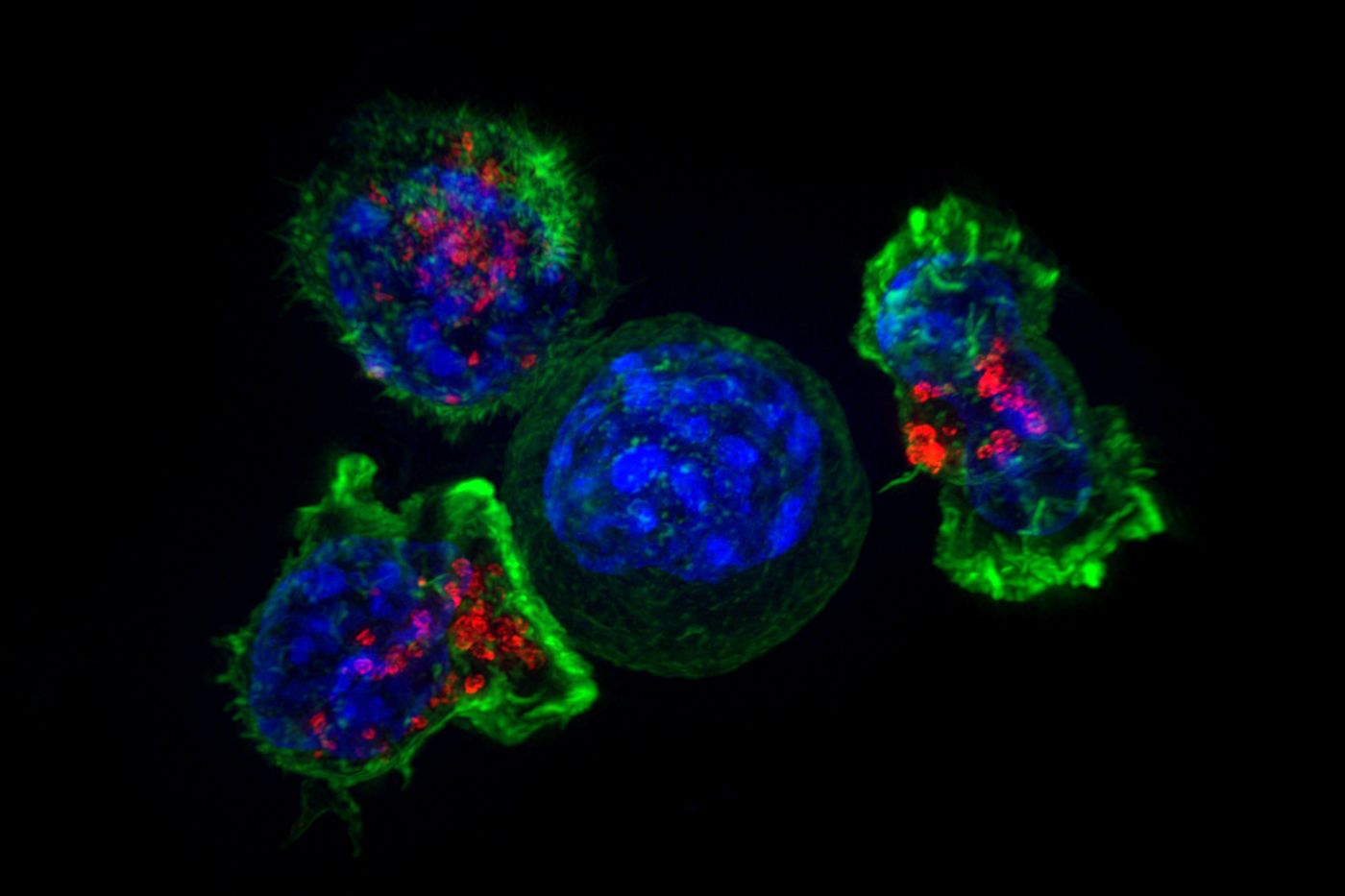
The immune system can detect and destroy pathogenic and cancerous cells, but sometimes those dangerous cells can evade the immune system. Researchers have now learned more about how cancer cells suppress the defensive function of killer T cells. New work has suggested that tumors release high levels of fat molecules into their local environment; when T cells take these fat molecules or oxidized lipids up, their cancer-killing ability is reduced. As they take up all that fat, the T cells increase the amount of CD36, a fat transporter, on their surface, which brings in even more oxidized fat, cycling the tumor-killing function down even further. The findings have been reported in Immunity.
"We know that tumors are a metabolically hostile environment for healthy cells, but elucidating which metabolic processes are altered and how this suppresses immune cell function is an important area of cancer research that is gaining a lot of attention," said senior study author Professor Susan Kaech, director of the Salk Institute's NOMIS Center for Immunobiology and Microbial Pathogenesis. "Our findings uncovered a novel mode of immunosuppression in tumors involving the import of oxidized fats in T cells via the cellular fat transporter CD36, which impairs their anti-tumor functions locally."
Tumor cells are known to absorb fat, and it's been thought that this increase in lipids disrupts immune function. This work can help researchers learn how fats are changing the tumor microenvironment. In cancer immunometabolism research, scientists are investigating the link between immune cell metabolism reprogramming in tumors and changes in nutrient availability.
In this study, the research team showed that tumor cells carry high levels of several types of lipids, especially oxidized lipids; these are often oxidized low-density lipoproteins (LDLs) that are considered to be 'bad' fat. When exposed to oxidized LDLs, killer T cells raise CD36 levels on their surface and take in more oxidized lipids. High levels of lipid oxidation in the cells disrupts their defensive action.
In a mouse model, the researchers found that CD36 caused T cells to take up lipids, which damaged the T cells and activated a stress response protein known as p38. "We found that when the T cells get stressed out by oxidized lipids, they shut down their anti-tumor functions," said first study author Shihao Xu, Ph.D., a Salk postdoctoral fellow.
This study also suggested that the function of T cells might be restored by blocking CD36 with antibodies or raising levels of a molecule that removes oxidized lipids from cells, called glutathione peroxidase 4 (GPX4).
Lipid oxidation also seems to happen in tumor cells (not just in T cells), and an excess of it can cause cells to die. Some researchers have suggested that it may be possible to kill tumor cells by increasing lipid oxidation, but Kaech and her team have urged caution.
"Now that we've uncovered this vulnerability of T cells to lipid oxidation stress, we may need to find more selective approaches to inducing lipid oxidation in the tumor cells but not in the T cells. Otherwise, we may destroy the anti-tumor T cells in the process, and our work shows a few interesting possibilities for how to do this," Kaech explained.
Experienced research scientist and technical expert with authorships on over 30 peer-reviewed publications, traveler to over 70 countries, published photographer and internationally-exhibited painter, volunteer trained in disaster-response, CPR and DV counseling.