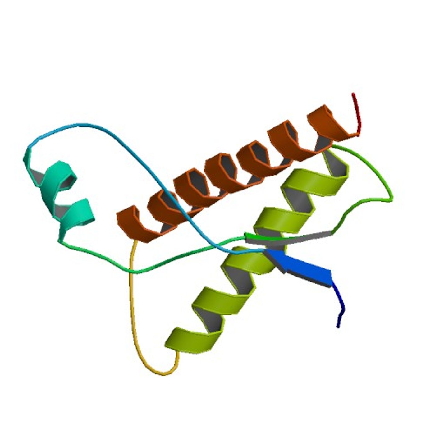
It is known that improperly folded proteins, called prion proteins, can cause serious detrimental health effects known as prion diseases. Examples include Creutzfeldt-Jakob disease and kuru in humans, mad cow disease in cows, scrapie in sheep and chronic wasting disease in deer. Understanding more about those conditions and developing therapeutics for them has been impeded by a lack of decent experimental models. New research described in The American Journal of Pathology outlines the different stages of prion disease in a mouse retina model. The investigators also describe an experimental model that can be exploited to test therapies. View the video below to learn more about prions and prion proteins.
"This work was done in collaboration with the USDA Agricultural Research Service and is an excellent example of how animal disease research can be leveraged to benefit human health," commented Heather West Greenlee, an Associate Professor in the Department of Biomedical Sciences at the Iowa State University College of Veterinary Medicine in Ames. "It provides important insights into the timeline for key pathologic milestones of prion disease in the retina and a model to study mechanisms of disease progression and evaluate therapeutic interventions."
It is thought that the development of transmissible spongiform encephalopathy (TSE) is dependent upon the movement of misfolded proteins from one structure in the central nervous system to another. To interrogate that process, the scientists injected scrapie (that was adapted for mice) into mouse brains and observed the spread of misfolded prion protein (PrPSc) via the optic nerve, from the brain to the retina until the onset of clinical disease, usually about 153 days post-inoculation (dpi).
The aim of the study was to characterize the temporal association between the transport of PrPSc from the brain to the retina, the buildup of PrPSc inside of the retina, the inflammatory response in the local retinal tissue, and the loss of neuronal cells.
The research team measured the time that passed between various stages of the disease progression as well as detecting sequential seeding of PrPSc in the retina at 60 dpi. After that, several more stages of disease development were identified: accumulation of PrPSc and retinal glia activation at 90 days, microglia activation at 105 dpi, then neuronal death in the retina at 120 dpi.
Prion diseases are also known as transmissible spongiform encephalopathies (TSEs); they are a transmissible protein misfolding disease, and as such make a good model for the study of common aspects of these disorders. The new model will also allow potential therapies to be evaluated according to the various issues that would need to be addressed - preventing accumulation of misfolded proteins, suppression of detrimental neuroinflammation, and neuron death prevention.
"Not only will this work contribute to the development of therapy to treat prion disease, but it may also provide important insights into which therapies may be effectively applied to the treatment of other protein misfolding diseases, including Parkinson disease and Alzheimer's disease," said Dr. Greenlee.
The video below is a long one, but at the beginning a patient with Creutzfeldt-Jakob disease briefly speaks about his condition. As the video continues, more information about the disease is presented.
Sources: AAAS/Eurekealert! via Elsevier, The American Journal of Pathology