For the first time, researchers have taken recordings of the behavior of cells in the epidermis as adult limbs regrow following amputation.
In new work published in the journal eLife, investigators from the Institute of Functional Genomics in Lyon (Institut de Génomique Fonctionnelle de Lyon, IGFL) in France captured continuous live imaging of a leg undergoing regeneration in a close relative of the common sand hopper, the crustacean Parhyale hawaiensis. That shellfish has been used as a model of limb regrowth since 2014, when scientists including Michalis Averof, Director of Research at the IGFL and senior author of the current study, introduced it.
"Parhyale hawaiensis is well suited for imaging limb regrowth. The animals have relatively rapid limb regeneration, requiring as little as one week for young adults to fully regrow their legs," Averof explained. "Also, their tiny limbs enable us to image the regeneration process in unprecedented cell-by-cell detail through their entire thickness." You can see the organism, and the collection of its embryos, in the video below.
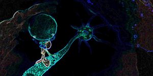
The research team labeled proteins in the epidermal cells of the limbs with fluorescence that would allow them to visualize and record the regeneration. Using microscopy, continuous recordings were made over the first few days of regrowth, during which the fluorescently labeled proteins were observable. Live imaging of the regenerating leg is shown below. Arrowheads are highlighting two apoptotic nuclei.
"Using this method, we identified a specific sequence of events and cell behaviors that unfold during limb regrowth," commented the first author of the report, Frederike Alwes.
"These include wound closure, followed by a quiet period when the epidermal cells migrate slowly towards the site of the wound, which then leads to extensive cell division and movement as the new leg starts to develop its shape. We were surprised to see that there was a sharp transition between the last two stages, which suggests the cells were coordinated by a common signal."
Additionally, the researchers determined that specialized stem cells are not required for the regrowing process. Most of the epidermal cells in the leg stump simply rearranged and divided, building up the new segments of leg.
"Traditionally, insight into cell behavior during limb regrowth has been gathered by imaging fixed samples and attempting to fill in the missing pieces, due to the difficulties in tracking cells during regeneration in active adult animals. With the ability to track the movements and behavior of single cells individually through time, we now have the means to understand the cellular dynamics of the regeneration process, which could not have been reconstructed from fixed material,” explained Alwes.
"While our paper focuses mostly on the behavior of epidermal cells, we now plan to extend this work to include all the different cell types that are involved in limb regrowth. The ultimate aim of our research is to explore how some animals can respond to a severe injury by regenerating an entire body part that was lost," Alwes concluded.
Sources:
AAAS/Eurekalert! via
eLifesciences.org/Agence Nationale de la Recherche,
eLife