The human brain contains everything we learn and all of the memories from our lives; the nerve cells in the brain and the molecular interactions between them function as the centers of that activity. To understand more about how we learn and how our brains hold memories, it's essential to understand the details of those connections between neurons, called synapses, and their chemical interactions through neurotransmitters, and how they change over time in a phenomenon known as synaptic plasticity. Scientists at Max Planck Florida Institute for Neuroscience (MPFI) in the lab of Ryohei Yasuda are using powerful microscopy tools to learn more. They have now published work in Neuron; they created molecular biosensors to reveal new characteristics of two proteins that have roles in synaptic plasticity, memory and learning, ERK and PKA. You can check out the video below from the researchers to learn more.
One part of a neuron that has been challenging to study by microscopy because it is so small is the dendritic spine. They are small protrusions on dendrites, which project in branches from neurons, and they have important roles in synaptic plasticity, can receive input from other neurons, and function in molecular signaling. Assaying for ERK and PKA activity works by checking for phosphorylation; they add a phosphate chemical group to a target protein. In dendritic spines, strong simulation strengthens and grows the spine, resulting in the formation of memories. Often a tool called western blotting is used to asses phosphorylation across a population of cells or in a tissue sample as an average; visualizing phosphorylation in dendritic spines directly has been difficult because they are so minuscule. Ada Tang, a postdoctoral researcher in Yasuda's lab, turned to biosensors to see the molecules.
She developed sREAChet, a new, dark molecule that absorbs light. She next linked sREAChet to both green fluorescent protein (GFP) and a peptide target of the protein and determined that it was able to indicate protein activity with a sensitivity that was 2-3 times better than previous sensors. The sensitivity was then good enough to use for visualizing the activity of individual dendritic spines.
"These sensors will be useful for researchers in a broad field of cell biology since ERK and PKA are involved in a variety of phenomena in cells and their abnormal activity is related to many diseases including cancer and mental diseases," Yasuda explained.
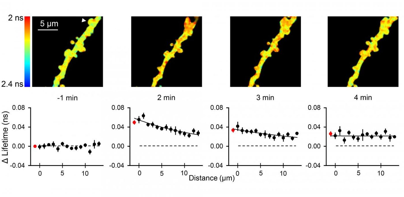
The researchers applied the new sensors to their work, using a 2-photon fluorescence lifetime microscope in the stimulation of single dendritic spines. They showed that ERK and PKA activity migrates from one spine. The investigators were surprised to find that the activity in the proteins was not confined to individual spines either, it spread over ten micrometers along the length of the dendrite, which influenced other dendritic spines in the area. It's estimated that the effect spreads up to tens of micrometers throughout a branch of dendrites. This team has already shown that the stimulation of only a few spines is enough to cause ERK activation in the nucleus of the neuron, but did not know the mechanism for that activation. They have now shown that when these proteins become activated within a spine, it's possible that the stimulation can spread over a long enough distance to reach the nucleus. "To find that PKA and ERK activation in spines is spreading for several tens of micrometers is certainly a surprising discovery for the field," said Tang.
This new tool to visualize activity may be an important step forward in the understanding of the biochemical properties of learning and memory.
Sources: AAAS/Eurekalert! via MPFI, Neuron