Reporting in Science, researchers have created an atlas that aims to show the location of proteins throughout a human cell. The work showed that many of these proteins, essential components of cells that are the functional readout of many coding genes, are located in multiple locations in any given type of cell. Led by Emma Lundberg, an Associate Professor at KTH Royal Institute of Technology in Stockholm, Sweden, this new data has been reported in Science.
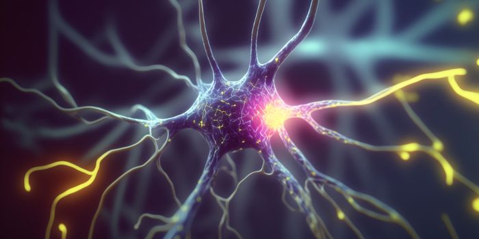
The research team has now outlined how proteins are distributed among the organelles and subcellular structures of a cell with unprecedented detail. Our bodies are made up of cells, and in order to carry out the myriad functions for life, cells have highly specialized roles that are dictated by our genomic blueprint. The specificity of cells is built upon the different proteins they express, as such knowing where proteins move or where they are stationed in a cell is an important part of understanding their function. Knowing more about protein function can only aid in our understanding of human disease.
This work was part of an international collaboration that created and analyzed more than 300,000 images, aiming to determine how the proteins of a human cell are organized and distributed. The researchers found protein location at single cell resolution, placing them to the compartments and structures within the cell.
The Cell Atlas is an open access tool that is free to investigators, and is the culmination of over a decade of work undertaken as part of the Human Protein Atlas program. The research report outlines the analysis of hundreds of thousands of images that were made for this effort, which also involved scientists in China, Denmark, Germany, South Korea and India.
"Only by studying the molecular components of the body's smallest functional unit, the cell, can we reach a full understanding of human biology," explained KTH Professor Mathias Uhlen, the Director of the Human Protein Atlas. "The Cell Atlas provides researchers with new knowledge that facilitates functional exploration of individual proteins and their role in human biology and disease."
Accompanying the research is a comparative work performed by Kathryn Lilley, Director of the Cambridge Centre for Proteomics at Cambridge University, UK, which enabled immunofluorescence (IF), a technique that uses antibody staining and microscopy analysis, to validate by an additional mapping tool that used mass spectrometry.
The investigators used 13,993 antibodies to sort 12,003 proteins into one or more of 30 different cellular compartments and organelles, thereby describing the protein makeup of 13 major organelles. Unsurprisingly, the nucleus and its substructures had the largest proteome, made up of 6,930 proteins, followed by the cytosol, with 4,279.
Intriguingly, nearly half of the proteins are not confined to one compartment, but instead appear in multiple places, suggesting that within a cell there is a pool of proteins that have roles in otherwise unrelated parts of the cell.
"The Atlas enables systems biology and cell modeling applications, and it is also a highly valuable resource for machine learning applications in image pattern recognition,” said Lundberg, who also oversees the High Content Microscopy facility at the Science for Life Laboratory (SciLifeLab).
Learn more about protein localization from the video above, a feature of MIT’s open courseware.
Sources: AAAS/Eurkealert! via KTH Royal Institute of Technology, Science