Our organs are made up of cells that are surrounded by a supportive matrix, which helps give the organ structure and form. The extracellular matrix exerts a significant influence on tissues and by extension, plays a role in disease progression. It has been very challenging to study it though, until now. Researchers at the Biotech Research & Innovation Centre (BRIC) at the University of Copenhagen have created a technique that reveals tissue and tumor structure in a new way.
Led by Professor Janine Erler, scientists have been able to dissolve away cells and leave the matrix intact. Investigators have revealed the inside of tumors and organs in a novel way and with unprecedented detail. The work has been reported in Nature Medicine.
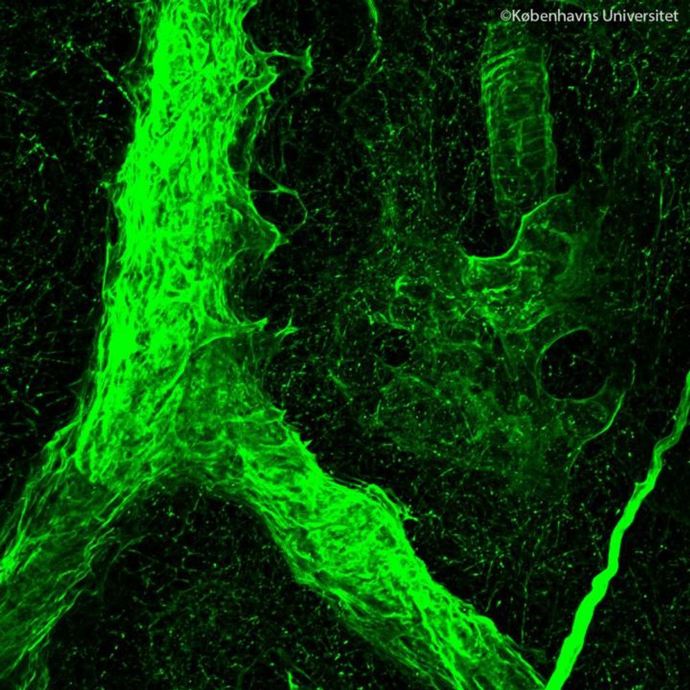
“We have developed a technique to obtain intact organ scaffolds and to image them in incredibly high detail using microscopes. We are the first to image the 3D structures of primary and metastatic tumors as well as healthy organs in this way,” said Erler.
The extracellular matrix provides a place for cells to anchor and arrange into an architecture. It also allows the cells inside to get a sense of changes in their environment and the appropriate responses. However, when there is dysfunction in this system, it can foster tumor growth. As such, scientists want to learn more about it, and this new tool is already providing new insights.
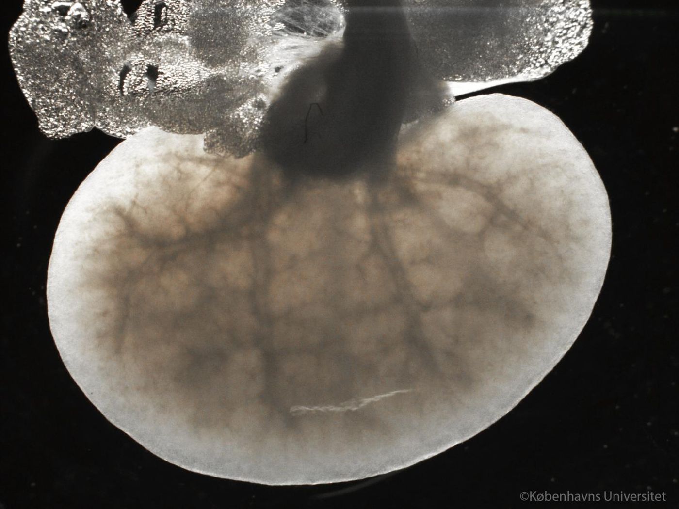
“We have isolated the structure that keeps tissues in place and organizes the cells inside them. We did this by using existing blood vessels to deliver cell-removing compounds directly to a specific tissue to remove all cells within an organ. Doing this leaves behind an intact scaffold that could be analyzed biochemically and microscopically, providing us with the first view of the structure of tumors,” explained Alejandro Mayorca-Guiliani, a postdoctoral fellow in Erler’s lab who pioneered the new method.
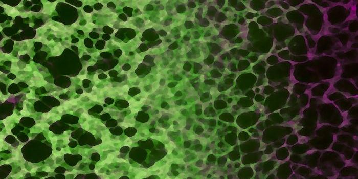
“When you remove the cells, the clarity of what you can see through the microscope is much improved – you can see the fibers of the matrix more clearly and you can look much deeper into the tissue. Using this approach, we have been able to see important differences in matrix organization when we looked at metastatic tumors in the lung and in the lymph node,” commented co-first author Chris Madsen, an imaging expert now working at Lund University in Sweden.
This work is sure to impact cancer research; viewing the remnants of organs stripped of their cells can illustrate how the architecture of organs and tumors differs.
“We are now re-introducing cells into our extracellular matrix scaffolds, bringing them back to life, to study how tumors form and how cancer progresses. This is extremely exciting and offers a unique opportunity to study how cells behave in their native environment,” concluded Professor Erler.
Learn more about the etracellular matrix from the video.
Sources: AAAS/Eurekalert! via University of Copenhagen, Nature Medicine