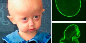
Scientists have found a way to highlight tumor tissue so it can be more effectively identified and removed. New work by researchers at the University of Pennsylvania School of Medicine has shown that surgeons can by find and excise cancerous nodules after they are made to glow. The new technique, which is described in the following video, has combined intraoperative molecular imaging (IMI) with a contrast dye that lights the cancerous tissue up by applying positron emission tomography (PET) scans before surgery. This work has been reported in the journal Annals of Surgery.
"Surgically removing tumors still leads to the best outcomes in cancer patients, and this study shows intraoperative molecular imaging can improve the surgeries themselves," noted first author Dr. Jarrod D. Predina, a post-doctoral research fellow in the Thoracic Surgery Research Laboratory and the Abramson Cancer Center's Center for Precision Surgery. "The more we can improve surgeries, the better the outcomes for these patients will be."
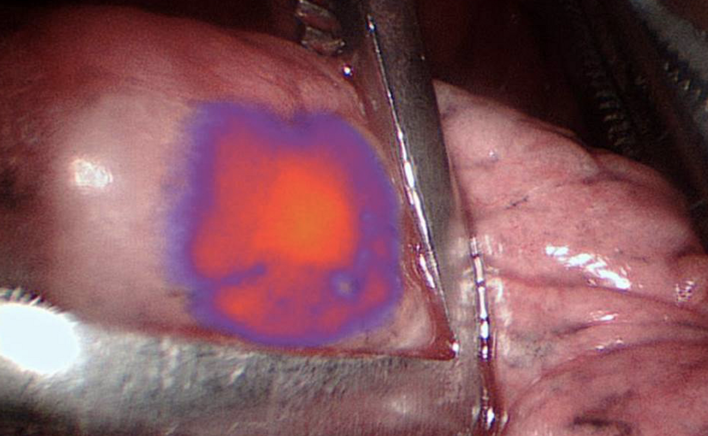
This research utilized OTL38, a contrast agent in the near-infrared spectrum. It can cause tumor cells to glow. Testing their technique, PET imaging was used along with IMI in 50 people that were having surgery to take out lung nodules. The pre-operative PET scans identified 66 nodules.
After applying the IMI, 91 percent of the nodules were found - 60 of 66. In addition, nine other nodules were revealed that had not been detected by the PET scan on its own.
Doctors identify pulmonary nodules, small growths in the lung, in around 250,000 Americans annually. Of those that are found, around 80,000 require removal because they might cause harm. PET scans are typically used prior to any procedure, and they find malignant nodules around 90 percent of the time. They obviously are not perfect. Tumors smaller than a centimeter can evade detection, and they can't tell the difference between cancer and benign growths. They also don' provide any information as the surgery happens. This new tool can change that.
"This shows the contrast agent is allowing us to remove more cancer from the patient than we would have with PET imaging alone," explained the senior author of the report, Dr. Sunil Singhal, the Director of the ACC's Center for Precision Surgery.
"PET imaging still has an important role to play in developing treatment plans for patients, but given its limitations, it's clear that IMI with this contrast agent can improve the picture surgeons are seeing," noted Dr. Singhal, who is the William Maul Measey Associate Professor in Surgical Research at Penn. "That's especially true when you're talking about nodules that are only a centimeter or smaller."
This work will hopefully improve outcomes for many people, and reduce the chances that patients will have to have additional future surgeries. One important thing will be getting this tool out there so that as many patients can benefit from it as possible.
Sources: UPI Via Penn Medicine, Annals of Surgery