Stem cells have so much promise because they can turn into virtually any type of cell in the body. Once, the only way to obtain these precious cells was by using fetal tissue, a process fraught with ethical complications. Then, a breakthrough technique enabled scientists to induce skin cells to turn into stem cells. Now researchers in many areas can use these special cells to learn more about human disease.
Researcher Aditi Deshpande, Ph.D. of the University of California San Francisco (UCSF) is utilizing this technique to perform research using skin cells from volunteers who carry a genetic feature linked to autism: the deletion or duplication of a region on chromosome 16. Then in the laboratory, those donated skin cells can be converted into stem cells, and then neurons after that. Learn more about this work from the video.
This particular region of chromosome 16 has been linked to both macrocephaly, an enlarged brain, as well as microcephaly, a small brain. There are 29 genes in that area of the chromosome. In 2009, Lauren Weiss, Ph.D., an Associate Professor of Neurology in the UCSF Department of Psychiatry and the UCSF Institute for Human Genetics connected this genetic region to brain disorders including autism and seizures, findings reported in the New England Journal of Medicine.
“What’s really interesting, is that although these subjects seem to have opposite features in terms of brain size, we see a related effect, based on whether they have fewer or more copies of the region,” explained Deshpande.
It is known that some models of autism suggest a link between the appearance or growth of a neuron and macrocephaly, she explained. “We wanted to know if the same thing is happening here.”
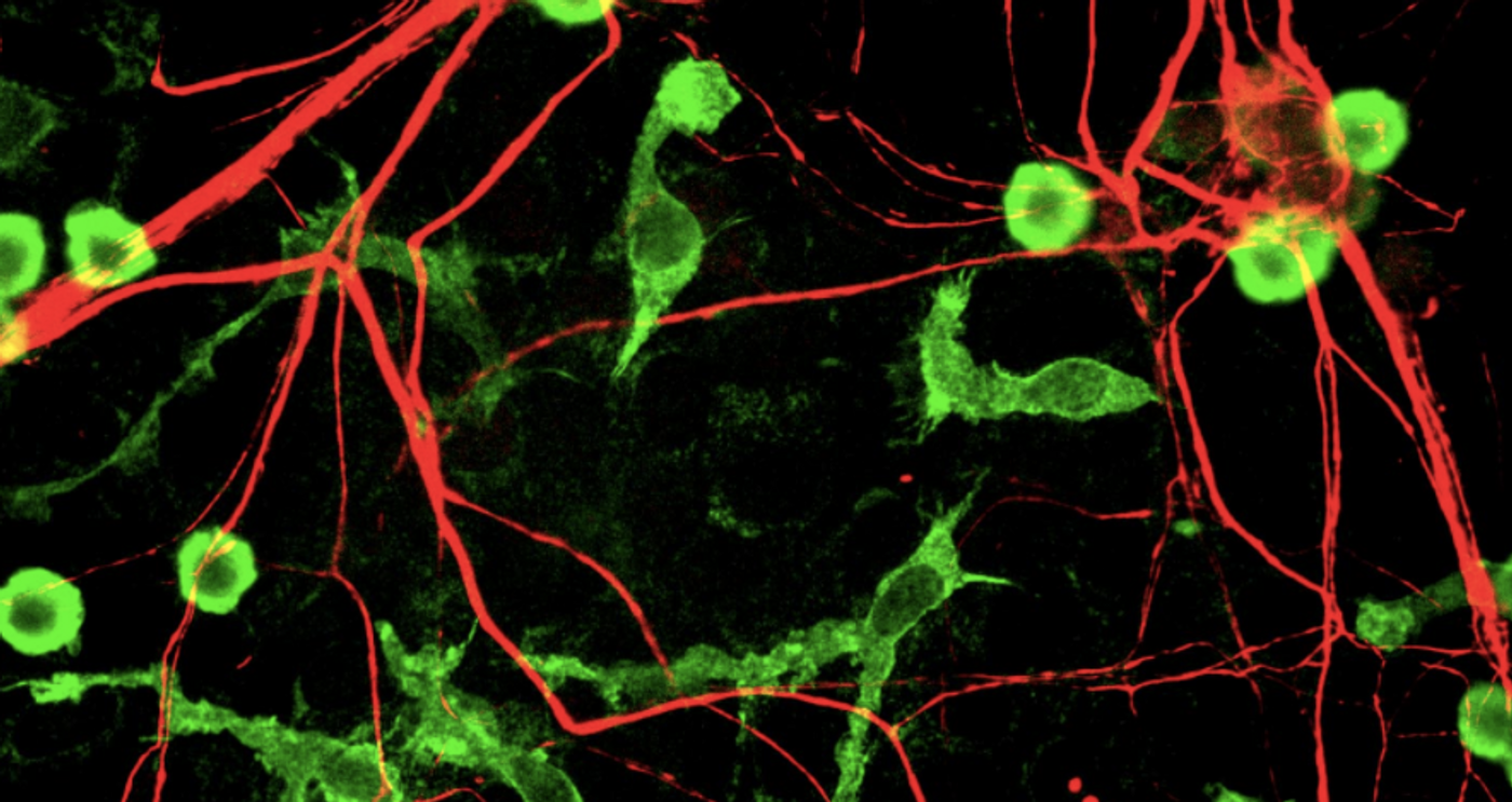
Deshpande assesses the neurons with a microscope. After staining the cells, software can count and analyze thousands of neurons that exhibit the various genetic deletions or duplications. She was able to detect differences between the aberrant neurons and normal ones.
Future work will investigate which of the 29 genes in the region are connected to the differences that are seen in the affected neurons. Deshpande is looking forward to this painstaking work.
“I simply love looking at neurons,” she said. “It really makes you appreciate the complexity of the brain.”
Source: UCSF