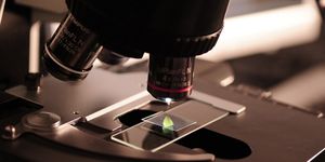
New research published in the journal Matter describes the development of a new kind of microscopy – this time at nanoscale. The research team says their method has the potential to revolutionize dispersion polymerization, a process that is used to make medicines, cosmetics, and latex on an industrial scale.
"Our method allows us to visualize this class of polymerization in real time, at the nanoscale, which has never been done before," said Northwestern's Nathan Gianneschi. "We now have the ability to see the reaction taking place, see these nanostructures being formed, and learn how to take advantage of the incredible things they can do."
Gianneschi, the associate director of the International Institute for Nanotechnology and the Jacob and Rosalind Cohn Professor of Chemistry in the Weinberg College of Arts and Sciences, in collaboration with Brent Sumerlin, the George and Josephine Butler Professor of Polymer Chemistry in the College of Liberal Arts & Sciences at the University of Florida, explain that nanoscale polymerization can make nanoparticles with desirable properties.
Nanoparticles are in high demand in industries ranging from environmental clean-up to “smart” drugs that deliver medicine directly into cells. Yet the process of producing and assembling nanoparticles has been delayed in part because the current technology does not allow scientists to see what is happening during the polymerization process.
To address this problem, the researchers figured out how to design a nanoscale polymer material that would initiate self-assembly inside of a liquid cell upon reaching a certain temperature. They were then able to record the resulting behavior of the block copolymers. They call their technique “variable-temperature liquid-cell transmission electron microscopy” (VC-LCTEM).
From this method, the researchers have begun to observe the process of self-assembly polymerization, noting the wide range of shapes that the nanoparticles take on. "These intricate and well-defined nanoparticles evolve over time, forming and then morphing as they grow," Sumerlin said. "What's incredible is that we're able to see both how and when these transitions occur in real time."

The research team has high hopes for the future of VC-LCTEM. “We think this can become a tool that's useful in structural biology and materials science too," said Gianneschi. "By integrating this with machine learning algorithms to analyze the images, and continuing to refine and improve the resolution, we're going to have a technique that can advance our understanding of polymerization at the nanoscale and guide the design of nanomaterials that can potentially transform medicine and the environment."
Sources: Matter, Eureka Alert