There is a new way to diagnose arthritis and watch its progression slowly over time to determine the best route of treatment. From the University of Cambridge, scientists introduce an algorithm based on three-dimensional imaging scans to monitor arthritis.
Unlike existing methods, the new approach can detect the smallest changes in arthritic joints. This way, scientists and physicians could test new treatments and understand more about how the disease develops. Learning more about arthritis could help experts better define the differences between severe cases and minor cases of arthritis.
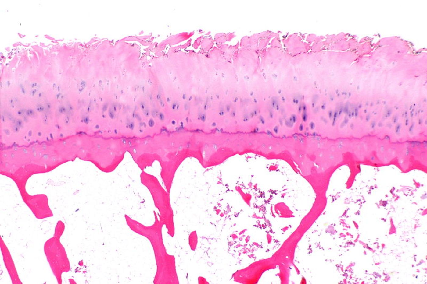
Osteoarthritis is a degenerative joint disease, the most common chronic condition of the joints. It develops when the cartilage that cushions joint movement deteriorates, causing stiffness, swelling, and pain. Excess weight, injury, and overuse can increase the risk of osteoarthritis, and it can only be treated with surgery to replace joints with an artificial alternative.
Current diagnostic methods for osteoarthritis include x-ray imaging, scans of which would show a loss of joint cartilage in joints between bones. However, this method requires human interpretation, which can vary, and it can’t effectively keep track of small changes in joint deterioration over time.
"Our ability to detect structural changes to identify disease early, monitor progression and predict treatment response is frustratingly limited by this,” said lead author Dr. Tom Turmezei.
The new technique is called joint space mapping (JSM), and it uses three-dimensional images from a standard computerized tomography (CT) scan. A CT scan produces cross-sectional images of anatomy, uses only low doses of radiation, and can be used by clinicians to diagnose and monitor various health conditions.
CT scan-directed JSM can successfully identify even the smallest changes in the space between the bones of an arthritic joint. Researchers conducted tests on human hip joints to develop an algorithm for diagnosing osteoarthritis with JSM, and they found that the new test is twice as sensitive – possibly more – as x-rays at detecting small structural changes in the joint
"Using this technique, we'll hopefully be able to identify osteoarthritis earlier, and look at potential treatments before it becomes debilitating," Turmezei explained. "It could be used to screen at-risk populations, such as those with known arthritis, previous joint injury, or elite athletes who are at risk of developing arthritis due to the continued strain placed on their joints."
The present study was published in the journal Scientific Reports.
Source: Arthritis Foundation, U.S. Food and Drug Administration, University of Cambridge