A super strong MRI approved by the U.S. Food and Drug Administration (FDA) may be releasing dangerous amounts of mercury from tooth fillings. From Akdeniz University in Turkey, scientists study the effects of the imaging technology to assess the risk of mercury poisoning.
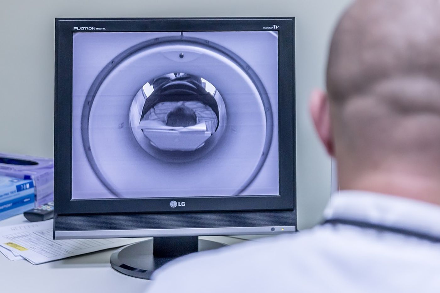
The ultra-high-strength 7-T MRI could be causing amalgam fillings in teeth to release toxic mercury. The 7-T was approved by the FDA in 2017, but used much less often than weaker MRIs, and it is not widely available. The MRI does improve sensitivity, contrast, and spectral dispersion. Lower strength MRIs, such as the more commonly used 1.5-Tesla (T) MRI or 3T MRI, don’t seem to carry the same risk of mercury poisoning. These imaging technologies are more common and often used for patient exams.
Amalgam (silver) fillings contain about 50 percent mercury, mixed with other metals like silver and tin. This filling material is the least expensive option for filling cavities caused by tooth decay. Amalgam fillings have been used for over a century in hundreds of millions of people, and they’re strong and long-lasting. These fillings are considered safe by the FDA for adults and children older than age six.
"In a completely hardened amalgam, approximately 48 hours after placing on teeth, mercury becomes attached to the chemical structure, and the surface of the filling is covered with an oxide film layer," explained lead author, Selmi Yilmaz, PhD. "Therefore, any mercury leakage is minimal."
Leakage may be more likely, though, under the influence of the 7-T MRI. Researchers from the new study set out to compare the amount of mercury released from a dental amalgam between 7-T and 1.5-T MRI scans. They placed extracted teeth with amalgam fillings in an artificial saliva solution which was exposed to either 1.5-T or 7-T. They compared these results to a control group placed in artificial saliva but exposed to neither MRI.
They found that the mercury content released by the teeth in the 7-T group was approximately four times the levels found in the 1.5-T group and the control group.
Yilmaz theorizes that this drastic disparity in mercury leakage is caused by “phase change in amalgam material or by formation of microcircuits, which leads to electrochemical corrosion, induced by the magnetic field."
For now, researchers know more studies are needed to understand how much of the mercury is absorbed by the body tissues to know what the health repercussions would be from this type of exposure.
The present study was published in the journal Radiology.
Sources: Stanford McNab Lab, Radiological Society of North America