When cells die, their genetic contents are released into bodily fluids through a process known as autolysis. In the recent past, scientists have exploited the presence of cell-free DNA in the blood to detect fetal DNA or tumor DNA. A
recent study goes even further to predict the origin of the cell-free DNA at the cellular level. This has implications for broadening the scope of detection for many health conditions, including metastatic cancer whose origins are sometimes unknown.
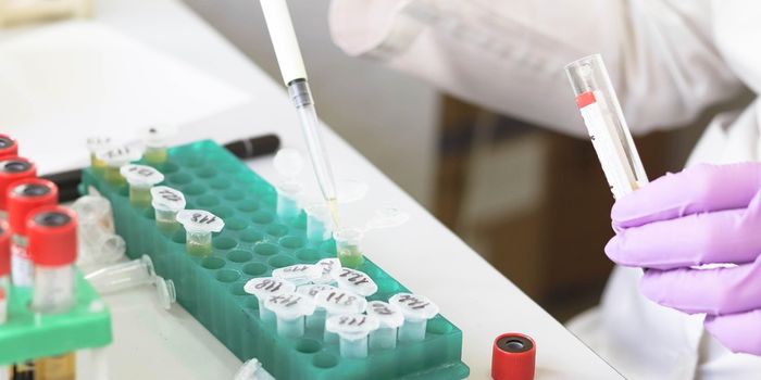
Traditional testing of cell-free DNA relies on comparing the DNA sequence between the patient’s healthy cells versus another DNA source. This source may be from a fetus, a tumor, or from a transplanted organ. However, these sequence-specific tests are limited, especially when no sequence variations are found.
Instead of relying on sequence variations, the
new method relies on the fragmentation patterns of the cell-free DNA.
In normal cells, DNA is wrapped around protein structures called the
nucleosomes, similar to how thread is wrapped around a spool. The spacing of the nucleosomes is unique to different cell types. As cells die, enzymes cut up free-floating DNA, specifically at sections that are not protected by the nucleosomes.
And because the pattern of nucleosome packaging is unique, so too are the fragmentation patterns of the cell-free DNA. This brilliant insight was discovered by Matthew W. Snyder, a graduate student in the laboratory of Dr. Jay Shendure, University of Washington professor of genome sciences and a Howard Hughes Medical Institute investigator.
Through deep sequencing of the cell-free DNA in blood, the researchers constructed maps of the nucleosome positions in the human genome. They essentially built an “address book” for the nucleosomes, from which they then used to trace the origins of various cell types.
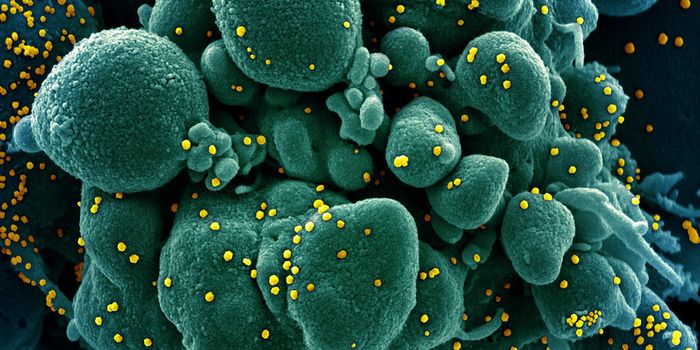
In healthy individuals, they found that the cell-free DNA had nucleosome patterns that correlated with normal blood cells. In cancer patients, however, the researchers found totally different nucleosome footprints in the cell-free DNA. By matching the nucleosome pattern in the patient’s blood to the nucleosome map, the scientists could identify the anatomical source of where the tumor originated.
"This could be particularly relevant in the 5 percent of metastatic cancers whose original source is unknown," Shendure said.
Because the new test doesn’t rely on sequence mismatches from different cells in the body, it can theoretically be applied to a broad range of health conditions that can’t be detected by traditional liquid biopsies. Heart attacks, strokes, and autoimmune diseases, for example, kill off certain cells and releases cell-free DNA with telltale nucleosome footprints. Knowing this can inform physicians on diagnosis and subsequent treatments.
Additional source:
MNT