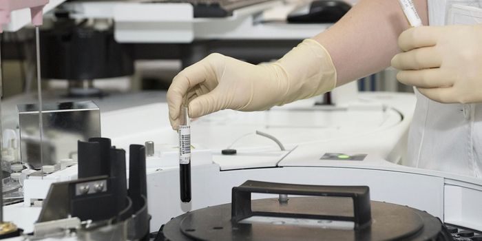
In recent years, more and more researchers are turning to nanoparticles for targeted delivery of drugs to select organs in the body. This technique promises to treat diseases, such as cancer, without the collateral damages to healthy cells. However, a recent study found that less than one percent of nanoparticle drug reach their intended target. To better visualize where nanoparticles really end up in the body, researchers at the University of Toronto are taking a “clear” approach – they’re making organs transparent.

Current methods of visualizing drug delivery are limited. Blood vessel walls, fat cells, and other biological barriers impede how much researchers can see inside an organ. “With current techniques, you can either see the whole organ, but without really knowing the details about what’s happening inside, or you can look at a single tissue slice in detail, but you lose contextual information,” said Shrey Sindhwani, MD-PhD candidate in the Institute of Biomaterials & Biomedical Engineering (IBBME), and first study author.
Stanford researchers studying the brain also reached this dilemma. Last year, they overcame the obstacle by developing a technique, known as CLARITY, which makes the brain transparent in order to visualize proteins with different antibodies.
Building on this existing method, the Toronto team applied it to other organs and nanoparticles. “People have used this technique to make tissues transparent, but we didn’t know it if would work with nanoparticles,” said Abdullah Syed, IBBME PhD candidate, and second author of the study.
To make the organs transparent, the team injected mouse organs and tissues with an acrylamide hydrogel. This substance bonds proteins together, and preserves the structural integrity of the organ. Lipid molecules, which are fat cells responsible for making organs opaque, are not linked together with the hydrogel. Thus, in subsequent washing steps, the lipids are easily removed, leaving organs that are virtually clear but structurally intact.
Nanoparticles injected into the organ were also held in place along with the cross-linked proteins. Thus once, the organ was clear, the researchers could image the nanoparticles in detail. Reportedly, this technique allowed the team to image nanoparticles at a depth of more than one millimeter, which is 25 times deeper than existing methods. In the liver, for example, “We see the nanoparticles stay near the vessel,” said Sindhwani. “Now that we know which cells are taking which nanoparticles up, we can appropriately target or deliver drugs for that cell type.”
“Our technique captures both perspectives with high resolution, producing a 3D map that can be zoomed in and out, like a Google Earth for the body,” said Sindhwani.
The team also reports that the process of making tissues and organs transparent can be optimized by tweaking the temperature and concentration of the detergent wash. “The original technique would take about a month to clear one sample,” said Syed. “We can do 48 tissues simultaneously in about 7 days.”
“The advantage of this process is that we could use the technique to study multiple diseases and conditions beyond cancer,” said Warren Chan, senior study author. “Nanoparticles have many potential applications in medicine, so we hope this technique will be adapted by researchers and clinicians in a number of different fields.”
Additional sources:
University of Toronto press release,
MNT