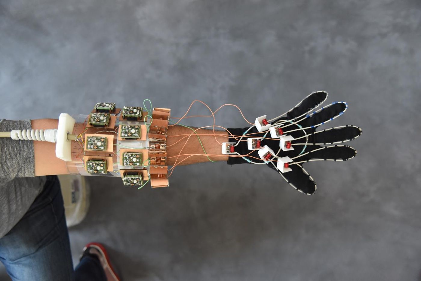
The newest in MRI technology literally fits like a glove. NYU School of Medicine scientists have designed an MRI glove that, for the first time, captures clear, high-quality images of bones, tendons, and ligaments in action.
Magnetic resonance imaging (MRI) technology came to life in the 1970s, and in the present study it’s being harnessed to visualize the interaction of different tissue types during movement. MRI works through a magnetic field that aligns hydrogen atoms in tissues of interest to create images from radio signals. Tissues emit these radio signals when they are “tipped” out of equilibrium by electromagnetic radiation.
"Our results represent the first demonstration of an MRI technology that is both flexible and sensitive enough to capture the complexity of soft-tissue mechanics in the hand," explained lead author Bei Zhang, PhD.
The diagnostic applications for this new technology are diverse and ground-breaking. First, the MRI glove could be used to diagnose repetitive strain injuries, such as carpal tunnel syndrome in office workers, athletes, and musicians. Scientists would also like to use the technology to create a “more versatile atlas of hand anatomy.” Information that this technology could uniquely provide would allow for more realistic hand models for guidance during surgical procedures. Additionally, scientists could use the technology to improve prosthetic design.
The key component of the glove design was constructing “high impedance” structures that remove interfering magnetic fields and provide clearer images of muscles, tendons, and ligaments in action. The new technology allows the visualization of fingers as they flex, and scientists can observe how different tissues in the hand interact with each other during different movements.
A simple comparison between a normal hand and an injured hand could provide scientists with detailed information about what happens in the tissue during injury.
"We wanted to try our new elements in an application that could never be done with traditional coils, and settled on an attempt to capture images with a glove," explained senior author Martijn Cloos, PhD. "We hope that this result ushers in a new era of MRI design, perhaps including flexible sleeve arrays around injured knees, or comfy beanies to study the developing brains of newborns."
The present study was published in the journal Nature Biomedical Engineering.
Sources: Radiologyinfo.org, NYU Langone Health/NYU School of Medicine