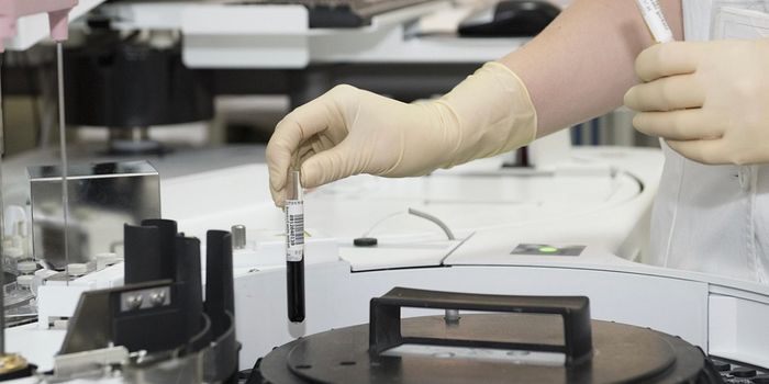
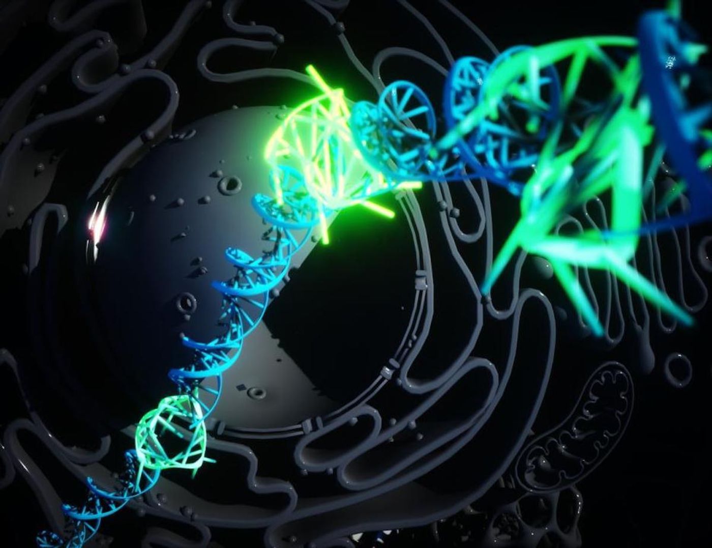
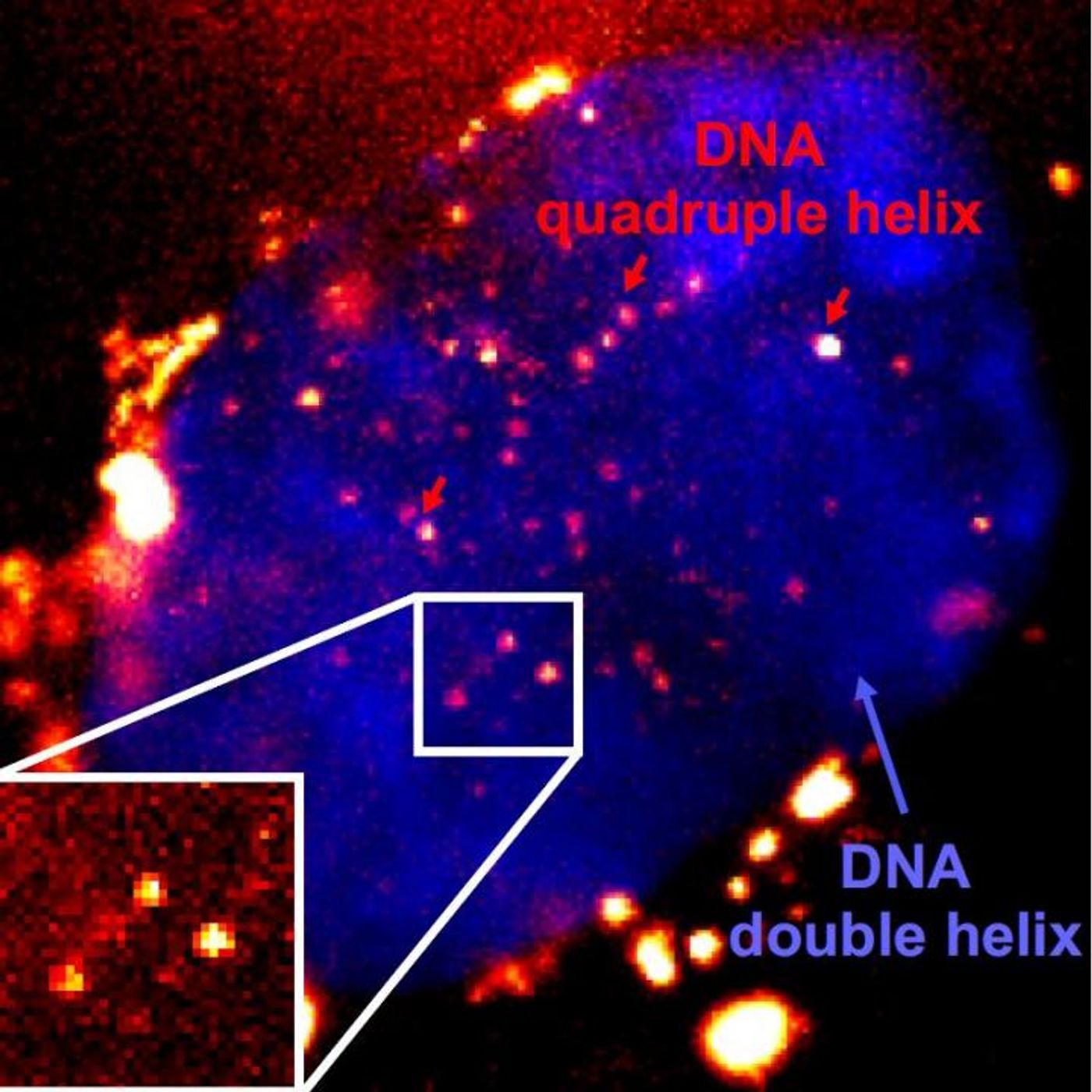
If you've seen a representation of a DNA molecule, you've seen the double helix, in which two strands of genetic material are wound together and linked by bonds. In the laboratory, scientists have gotten DNA to form other structures, including a quadruple helix, called DNA G-quadruplexes (G4s). Now researchers have created a fluorescent marker that can be used to bind to G4s so that they can be visualized. The findings have been reported in Nature Chemistry.
"For the first time, we have been able to prove the quadruple helix DNA exists in our cells as a stable structure created by normal cellular processes. This forces us to rethink the biology of DNA. It is a new area of fundamental biology, and could open up new avenues in diagnosis and therapy of diseases like cancer," said one of the study authors, Dr. Marco Di Antonio, now of Imperial College London. "Now we can track G4s in real-time in cells. We can ask directly what their biological role is. We know it appears to be more prevalent in cancer cells and now we can probe what role it is playing and potentially how to block it, potentially devising new therapies."
The researchers suggested that portions of DNA will form G4s to briefly hold open their structure, potentially to facilitate processes like transcription, in which the genetic code is read and the instructions for making a protein are transcribed from DNA into RNA. G4s have also been associated with genes that are linked to cancer and have been found at high levels in cancerous cells.
In this work, the researchers were able to reduce the levels of G4-detecting molecules that were being used, so that the detection process isn't interfering with and disrupting the DNA in the cell.
"Scientists need special probes to see molecules within living cells, however, these probes can sometimes interact with the object we are trying to see," explained study author Dr. Aleks Ponjavic, now of the University of Leeds. "By using single-molecule microscopy, we can observe probes at 1000-fold lower concentrations than previously used. In this case, our probe binds to the G4 for just milliseconds without affecting its stability, which allows us to study G4 behavior in their natural environment without external influence."
The new G4-detecting probe employs an intense fluorescent molecule that can easily stick to G4 structures even at very low levels. Not every G4 in a cell is marked, but single G4s can be seen at a high enough resolution to track them individually.
The researchers used their probe to reveal that G4s seem to rapidly appear and dissipate, which may suggest that their function is specific, and the cell might have to carefully control it to avoid damage.
Experienced research scientist and technical expert with authorships on over 30 peer-reviewed publications, traveler to over 70 countries, published photographer and internationally-exhibited painter, volunteer trained in disaster-response, CPR and DV counseling.

![WGS for rare disease diagnosis [eBook]](https://d3bkbkx82g74b8.cloudfront.net/eyJidWNrZXQiOiJsYWJyb290cy1pbWFnZXMiLCJrZXkiOiJjb250ZW50X2FydGljbGVfcHJvZmlsZV9pbWFnZV84MmRlM2UyYjA5M2Q3ZTYwOTI3Zjc1YTRjOWU2N2RmMjkzMThjMTJkXzI1MDcucG5nIiwiZWRpdHMiOnsidG9Gb3JtYXQiOiJqcGciLCJyZXNpemUiOnsid2lkdGgiOjcwMCwiaGVpZ2h0IjozNTAsImZpdCI6ImNvdmVyIiwicG9zaXRpb24iOiJjZW50ZXIiLCJiYWNrZ3JvdW5kIjoiI2ZmZiJ9LCJmbGF0dGVuIjp7ImJhY2tncm91bmQiOiIjZmZmIn19fQ==)