According to new research, computed tomography (CT) scans can successfully detect fractures using 14-times less the amount of radiation than a standard CT scan. The findings were presented today, March 2, at the 2016 American Academy of Orthopedic Surgeons (AAOS) Annual Meeting.
CT scans are the most frequently-used imaging tools in medicine. Doctors consider the overall risk low, but radiation has been linked to an increased risk of cancer. The issue has raised public health concerns regarding how the radiation exposure affects children and young adults. About 600,000 head and abdominal CT scans are performed each year on children under 15. Federal agencies and medical societies are working to reduce the number of unnecessary scans.
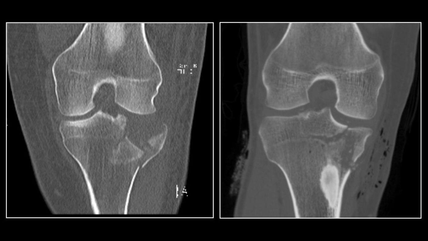
CT scans take x-ray images at different angles and combine them to produce virtual "slices" of specific areas of the object. They are used to help diagnose conditions such as cancers, internal bleeding, or signs of heart disease. In orthopaedic trauma, clinicians use CT scans to help diagnose broken bones and injuries around a joint. The scans also help doctors see where a joint is displaced. This helps in determining how to put the joint back into place.
Radiologist Soterios Gyftopoulos led a team of researchers in finding how to reduce the amount of radiation delivered by a CT scan without compromising the image. The NYU Langone Medical Center researchers established a protocol called REDUCTION (Reduced Effective Dose Using Computed Tomography In Orthopaedic Injury). The protocol was proven effective in a previous study that used the protocol to examine air around knee joints.
The study recruited 50 participants who, between August 2014 and March 2015, showed clinical symptoms of joint fractures. They received ultra-low dose radiation CT scans. The researchers compared the participants' scans with standard CT scans of patients who were the same age and had similar fracture injuries.
They found the ultra-low dose scans detect joint fractures 98 percent of the time. Orthopaedic surgeons rated the image quality as moderate to near perfect. Ultra-low dose CT scans achieved 98-percent sensitivity and 89 percent specificity. Standard scans achieve 98 percent sensitivity and 85 percent specificity with occult fractures removed.
"The ability to perform ultra-low dose radiation CT scans without compromising image quality demonstrates the comprehensive capabilities of this protocol," says senior study author Kenneth A. Egol, chief of the division of orthopaedic trauma surgery at NYU Langone. "Patients who undergo a traumatic injury or suspected fracture have enough to worry about. Our research makes radiation exposure among the least of their concerns."
The researchers are conducting follow-up on the patients to find whether the outcome of the finding changes.
Sources:
NYU Langone Medical Center press release via EurekAlert!