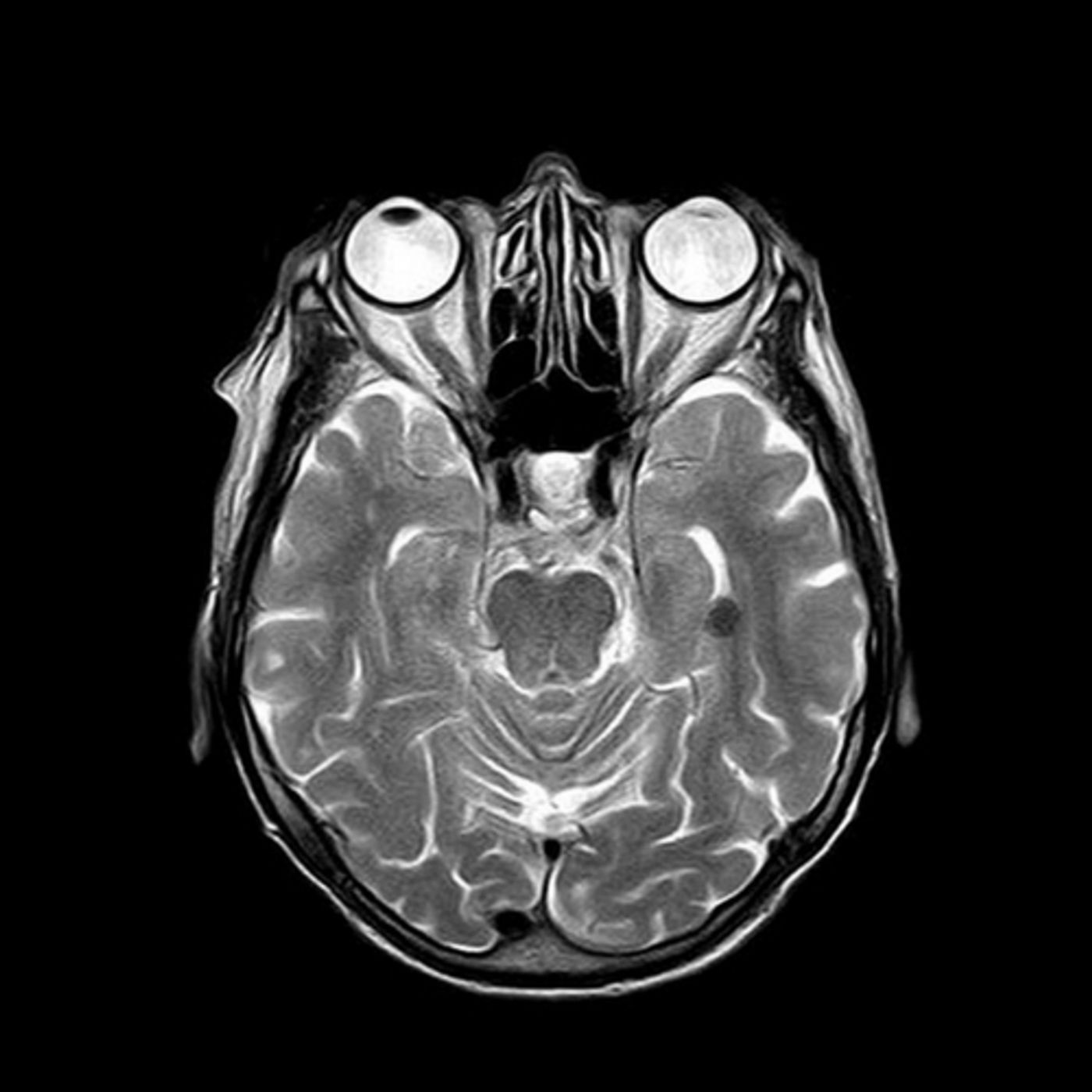
MRI scans can look deep into the brain using powerful magnets to see brain anatomy and structure. Different types of MRI scans including functional MRIs that track blood flow as an indicator of brain activity in real time, and diffuse tension MRIs that image brain function by following water molecules in the brain are just two examples of specific MRI scans that go beyond the typical.
Researchers at MIT have now developed technology that can look directly at neurons and their firing patterns. It's done by having an MRI scan that can detect calcium ions. Calcium ions are involved directly in patterns of neural activity in the brain. Rather than looking at blood flow or water deposits, the new type of scan hones in on calcium ions, and it's this activity that could reveal how different regions of the brain communicate.
Alan Jasanoff is an MIT professor of biological engineering, brain, and cognitive sciences, and nuclear science and engineering, and also an associate member of MIT's McGovern Institute for Brain Research. He led the research into this technology and explained, "Concentrations of calcium ions are closely correlated with signaling events in the nervous system. We designed a probe with a molecular architecture that can sense relatively subtle changes in extracellular calcium that are correlated with neural activity."
The team at MIT showed that their calcium sensor accurately detected changes in neural activity in an area deep in the brain called the striatum. They used lab rats and induced changes in the neural firing patterns by electrical stimulation and chemical injections. The sensor, which will be used in the MRI scans picked up even small changes in calcium that indicated brain activity.
The gold standard of neuroscience research, MRI scans let scientists and medical professionals see what parts of the brain are active in what kinds of tasks. Functional MRIs look at blood flow, but there isn't a direct relationship between blood flow to one area and specific brain tasks. Calcium ions are different. They flow directly into brain cells when a neuron fires. Typically, scientists use fluorescent molecules to bind to calcium to make them show up on microscopy studies, but this is only doable on small sections of the brain.
The team at MIT developed an MRI sensor that uses two particles that bind to calcium. These particles then cluster together with the calcium ions and appear darker on MRI images. There's an inverse relationship in the process meaning that high levels of calcium outside of cells corresponds to low neuron activity. When calcium ions drop, that indicates neurons are firing.
The sensors picked up these increases and drops in calcium in lab rats, so the team hopes to move forward with further research. One of the goals is to find a way to get the particles that bind to calcium ions to pass through the blood-brain-barrier so that they could be administered to larger areas of the brain. The technique shows great promise in helping researchers map brain activity. Known the mechanism in the brain of some diseases and conditions can help point scientists in the right direction in treating patients. The video below is from MIT and explains in more detail how the imaging works, check it out.
Sources: Nature Nanotechnology MIT.